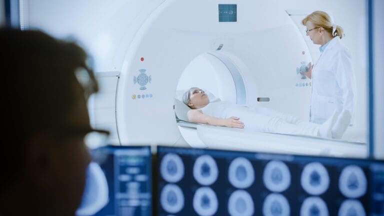
Understanding brain lesions in multiple sclerosis (MS) on MRI is key for correct diagnosis and treatment. At Liv Hospital, we use the latest MRI tech to spot and study these lesions. This gives us important info on MS’s progress and how well treatments work.
MS brain lesions show up as bright white spots on T2-weighted MRI images. They might also look dark or black on T1-weighted images. Studies, like those on Medical News Today, show that the number and type of lesions can tell us how severe the condition is. This helps doctors make better treatment choices.
Key Takeaways
- MRI is a critical tool for diagnosing and managing multiple sclerosis.
- MS brain lesions appear differently on various MRI sequences.
- The number and type of lesions can indicate disease severity.
- Liv Hospital uses advanced MRI technology for accurate diagnosis.
- Effective management of MS relies on precise lesion analysis.
Understanding Multiple Sclerosis and Brain Lesions
Multiple sclerosis (MS) is a chronic disease that affects the central nervous system. It’s an autoimmune disease that can be disabling. To understand MS, we need to know how it causes brain lesions.
What is Multiple Sclerosis?
MS happens when the immune system attacks the myelin, the protective covering of nerves. This disrupts communication between the brain and the body. It leads to various neurological symptoms, depending on where the damage is.
How MS Affects the Brain
MS damages the brain in different ways, mainly through lesions. These lesions can be in white matter, gray matter, or optic nerves. Each person’s case is unique, thanks to the varied locations and types of lesions.
Advanced imaging, like MRI, is key in spotting and tracking these lesions.
The Role of Demyelination in Lesion Formation
Demyelination is the damage to the myelin sheath. This damage disrupts nerve signals, causing MS symptoms. The extent of demyelination and repair efforts affect the disease’s progression.
MS lesions often appear in specific areas like the brainstem and spinal cord. Studies on conditions like prediabetes show the value of MRI in diagnosing MS.
MRI as the Gold Standard for MS Diagnosis
MRI is the top choice for diagnosing Multiple Sclerosis (MS). It gives us detailed brain images. This helps doctors spot and track MS with great accuracy.
Thanks to MRI, we can see lesions and understand how the disease is progressing. This has changed how we approach MS care.
Types of MRI Sequences Used in MS
There are different MRI sequences for MS diagnosis. These include T1-weighted, T2-weighted, and contrast-enhanced images. Each type shows different things about the lesions.
T1-weighted images are great for seeing the brain’s structure. They help find lesions that look darker, or “black holes.” T2-weighted images are better at spotting lesions. They show up brighter, helping to count how many lesions there are.
T1-Weighted vs. T2-Weighted Images
Knowing the difference between T1 and T2 images is key in MS diagnosis. T1 images show chronic damage and severe lesions. T2 images give a wider view of all lesions and activity.
| MRI Sequence | Lesion Appearance | Clinical Utility |
| T1-Weighted | Hypointense (“Black Holes”) | Assesses chronic damage |
| T2-Weighted | Hyperintense | Evaluates total lesion load and activity |
The Importance of Contrast Enhancement
Contrast enhancement with gadolinium is key for finding active lesions. Active lesions show the disease is active. This is important for diagnosing and tracking MS.
Contrast enhancement helps doctors tell the difference between new and old lesions. This is vital for understanding the disease’s progress.
What Do Lesions on the Brain Look Like on MRI?
Brain lesions on MRI are key for diagnosing Multiple Sclerosis. They show how severe the disease is. Lesions can look different based on the MRI sequence used.
Hyperintense Lesions on T2-Weighted Images
On T2-weighted MRI images, MS lesions show up as hyperintense or bright white spots. These spots mean the myelin sheath is damaged. This damage causes inflammation and swelling.
These bright spots are often found in the brain’s white matter. They are most common near the ventricles. The number and location of these spots tell us a lot about the disease.
Hypointense “Black Holes” on T1-Weighted Images
On T1-weighted images, some MS lesions look hypointense or dark. These are called “black holes.” They show more severe damage and loss of nerve fibers.
Black holes can mean a worse prognosis and more disability. Not all dark spots are permanent. Some may light up with gadolinium, showing active inflammation.
Gadolinium-Enhancing Active Lesions
Gadolinium contrast in MRI scans helps spot active MS lesions. Gadolinium-enhancing lesions are bright on T1-weighted images. They show where the blood-brain barrier is broken and there’s active inflammation.
These active lesions are important for checking disease activity and treatment success. The number and where they are can help decide treatment and show how the disease is progressing.
Characteristic Locations of MS Brain Lesions
Knowing where MS brain lesions occur is key for diagnosis and treatment. These lesions don’t spread out randomly. They often show up in certain parts of the central nervous system.
Periventricular Lesions
Lesions around the ventricles, fluid-filled spaces in the brain, are common in MS. Periventricular lesions are seen on MRI scans as bright spots. They are important because they can show how the disease is progressing.
Juxtacortical and Cortical Lesions
Lesions near or in the cortex, the brain’s outer layer, are typical of MS. Juxtacortical lesions are next to the cortex, and intracortical lesions are inside it. These can be harder to spot but are vital for understanding the disease’s spread.
Corpus Callosum Involvement
The corpus callosum, a thick band of nerve fibers, is another common site for MS lesions. Lesions here are often a sign of MS, as they’re rare in other conditions.
Infratentorial and Spinal Cord Lesions
MS lesions can also appear in the infratentorial region, which includes the cerebellum and brainstem, and in the spinal cord. These spots are important because they can cause a variety of symptoms. Lesions here can affect coordination, balance, and how the body works on its own.
The places where MS brain lesions appear are key for diagnosing and managing the disease. By knowing where these lesions are likely to be, doctors can better read MRI scans and plan treatments.
MS Lesions vs. Normal Brain Appearance on MRI
It’s key to know the difference between MS lesions and a normal brain on MRI for correct diagnosis. When looking at MRI scans, we must tell apart the changes seen in Multiple Sclerosis from those in a healthy brain.
Normal Brain MRI Appearance
A normal brain MRI shows clear structures without big problems. The white matter looks bright on T1-weighted images, and the gray matter is darker. The ventricles and sulci are even, and there are no signs of lesions or abnormal enhancement.
Age-Related White Matter Changes vs. MS Lesions
Age-related white matter changes can look like MS lesions on MRI. But, there are important differences. Age-related changes are more spread out and less clear, often in the subcortical white matter. MS lesions, on the other hand, are more focused and often near the ventricles.
Key Differentiating Features
There are several ways to tell MS lesions apart from normal age-related changes or other conditions. These include:
- Location: MS lesions often appear in the periventricular, juxtacortical, and infratentorial areas.
- Shape and Size: MS lesions can differ in size and shape but are usually ovoid and go straight across the ventricles.
- Enhancement: Active MS lesions might show up on post-contrast T1-weighted images.
| Feature | MS Lesions | Age-Related Changes |
| Location | Periventricular, juxtacortical, infratentorial | Subcortical white matter |
| Appearance | Focal, ovoid lesions | Diffuse, less distinct |
| Enhancement | May enhance with contrast | Typically does not enhance |
Knowing these differences helps doctors better diagnose and treat MS. They can tell it apart from other conditions that might look similar on MRI.
How Many Brain Lesions Are Normal in MS?
The number of brain lesions in MS can vary a lot among patients. MS is a chronic disease where the body attacks the protective covering of nerve fibers in the brain and spinal cord. The number and presence of brain lesions are key in diagnosing and tracking the disease.
Lesion Load and MS Diagnosis
Lesion load is the total number and size of lesions seen on MRI scans. A higher lesion load often means a more severe disease. But, the link between lesion load and symptoms is not always clear. Some patients with many lesions may have few symptoms, while others with fewer lesions may have severe symptoms.
We use MRI to count lesion load and track changes over time. This helps us understand how the disease is progressing and make better treatment choices.
The McDonald Criteria for MS Diagnosis
The McDonald criteria are guidelines for diagnosing MS. They consider clinical symptoms, MRI findings, and other tests to confirm an MS diagnosis. The 2017 update of the McDonald criteria highlights the role of MRI in showing lesions in different parts of the brain over time.
Key components of the McDonald criteria include:
- Clinical attacks
- Lesion dissemination in space (DIS)
- Lesion dissemination in time (DIT)
- MRI findings
Significance of Lesion Number and Distribution
The number and where lesions are found are important in diagnosing MS and understanding its severity. Lesions can appear in different parts of the CNS, like the brain, spinal cord, and optic nerves.
| Lesion Location | Clinical Significance |
| Periventricular | Common in MS, often associated with cognitive symptoms |
| Juxtacortical and Cortical | May be associated with cognitive and motor symptoms |
| Infratentorial | Can cause coordination and balance problems |
Knowing the importance of lesion number and location helps us create personalized treatment plans for each patient.
Correlation Between MS Lesion Location and Symptoms
Understanding how MS lesions affect symptoms is key to managing the disease. MS is different for everyone, and symptoms vary due to where lesions are in the brain. This knowledge helps doctors tailor treatments.
The location of MS lesions in the brain greatly affects symptoms. For example, lesions near the optic nerve can cause vision problems. This shows how specific symptoms can be tied to certain lesion locations.
Visual Symptoms and Optic Nerve Lesions
Optic nerve lesions can lead to vision issues like blurred or double vision. Inflammation of the optic nerve, known as optic neuritis, is common in MS. It can severely impair vision.
Motor Symptoms and Corticospinal Tract Lesions
Lesions in the corticospinal tracts can cause weakness and coordination problems. These symptoms can greatly affect a person’s ability to move and live their life.
Sensory Symptoms and Sensory Pathway Lesions
Sensory pathway lesions can cause numbness, tingling, and pain. These symptoms can be very distressing and impact a person’s overall health.
Cognitive Symptoms and Cerebral Lesions
Cerebral lesions can lead to memory and attention problems. These cognitive issues can make everyday tasks hard and reduce independence.
Knowing how MS lesions relate to symptoms helps doctors predict outcomes. This knowledge allows for more effective treatment plans to manage symptoms.
Advanced MRI Techniques for MS Lesion Detection
Advanced MRI techniques have greatly improved how we find and understand MS lesions. They give us new insights into how the disease progresses. This makes diagnosing MS more accurate.
FLAIR and DIR Imaging
Fluid-attenuated inversion recovery (FLAIR) imaging is great for spotting brain lesions. It’s good at finding lesions in certain areas of the brain. FLAIR makes these lesions stand out by hiding the signal from cerebrospinal fluid (CSF).
Double inversion recovery (DIR) imaging takes it a step further. It hides signals from both CSF and white matter. This makes cortical lesions even clearer.
These methods are key for spotting MS lesions. They help doctors diagnose and keep track of the disease.
Diffusion Tensor Imaging
Diffusion tensor imaging (DTI) shows how well white matter tracts in the brain are doing. It looks at how water molecules move. This helps spot small changes in tissue structure, which is important for seeing MS damage.
Magnetization Transfer Imaging
Magnetization transfer imaging (MTI) is good at finding demyelination and axonal loss. These are common in MS. MTI gives numbers that show how much damage there is. This helps doctors see how the disease is changing and how treatments are working.
Susceptibility-Weighted Imaging
Susceptibility-weighted imaging (SWI) is great at finding iron deposits and other substances in MS lesions. It can spot lesions that other MRI sequences can’t. This gives a fuller picture of the disease’s impact.
The table below shows the advanced MRI techniques we talked about. It explains how they help find MS lesions:
| MRI Technique | Application in MS |
| FLAIR Imaging | Detection of periventricular and juxtacortical lesions |
| DIR Imaging | Enhanced visibility of cortical lesions |
| DTI | Assessment of white matter tract integrity |
| MTI | Quantification of demyelination and axonal loss |
| SWI | Detection of iron deposits and other substances in lesions |
Conclusion: The Significance of Brain Lesions in MS Management
Understanding brain lesions in multiple sclerosis (MS) is key to managing the condition well. MRI is vital for diagnosing and tracking MS. It shows the presence and details of ms lesions on the brain.
MRI’s role in MS diagnosis is clear. It uses T1-weighted and T2-weighted images to give insights into brain lesions. This helps doctors decide the best treatment plan.
It’s important to know about ms lesions on the brain and their effects on patients. The location of ms brain lesions and symptoms are linked. Advanced MRI techniques help manage MS better.
By learning more about brain lesions in MS and using MRI, we can improve patient care. This leads to better health outcomes and top-notch healthcare for international patients.
FAQ
What do MS lesions look like on MRI?
MS lesions show up as bright spots on T2-weighted images. They appear as dark “black holes” on T1-weighted images. Active lesions can also be seen with gadolinium enhancement on MRI.
How many brain lesions are normal with MS?
The number of brain lesions in MS varies a lot. The McDonald criteria look at the number and where the lesions are. But, there’s no one “normal” number of lesions.
What does MS look like on the brain?
MS lesions are found in specific areas. These include periventricular, juxtacortical, and cortical areas. They are also found in the corpus callosum and in the spinal cord.
How does MS affect the brain?
MS causes demyelination, leading to lesions. These lesions can disrupt brain function. This disruption causes a variety of symptoms.
What is the difference between MS lesions and age-related white matter changes?
MS lesions are more common and widespread than age-related changes. They are also found in specific areas, like the periventricular region.
Can lesions on the brain go away?
Some MS lesions may get smaller or disappear over time. But, others stay the same. Treatments can help reduce the number and size of lesions.
What are MS lesions on the brain?
MS lesions are damaged areas in the brain due to demyelination. They can be seen on MRI and are a key sign of MS.
How are MS lesions detected on MRI?
MRI uses T1-weighted and T2-weighted images to find MS lesions. Gadolinium contrast is also used to enhance detection.
What is the significance of lesion location and symptoms in MS?
The location of MS lesions can relate to specific symptoms. This includes visual, motor, sensory, and cognitive symptoms. Knowing these connections helps in managing MS.
What advanced MRI techniques are used to detect MS lesions?
Advanced MRI techniques include FLAIR, DIR imaging, and diffusion tensor imaging. Magnetization transfer imaging and susceptibility-weighted imaging are also used. These help detect and understand MS lesions better.
References:
- Medical News Today. https://www.medicalnewstoday.com/articles/323976
- Brain lesions ScienceDirect. https://www.sciencedirect.com/topics/neuroscience/brain-lesion


