Last Updated on November 27, 2025 by Bilal Hasdemir
Do MRI Show Broken Bones
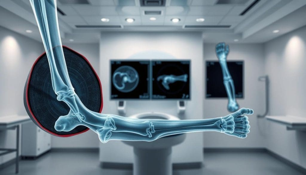
Diagnosing broken bones needs clear and confident imaging. At Liv Hospital, advanced tech and care for patients come together. Knowing how MRI compares to X-ray helps patients make smart health choices.
X-rays are often the first tool used to check for fractures. But MRI scans are better at showing soft tissues. This includes muscles, blood vessels, ligaments, and organs. They’re great for finding small or hidden breaks.
Key Takeaways
- MRI is great for finding small or hidden fractures.
- X-rays are usually the first tool used.
- MRI scans are better at showing soft tissues.
- MRI can find fractures where X-rays can’t.
- Liv Hospital uses the latest tech and cares for patients.
The Fundamentals of Bone Imaging
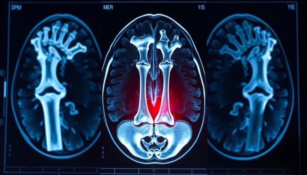
Medical imaging has changed how we find and treat bone fractures. It gives us clear and precise views of bones. This is key for good patient care. Many imaging methods have been created to help see bone structures well.
The Evolution of Medical Imaging for Fracture Detection
Diagnosing bone fractures has improved a lot. Wilhelm Conrad Röntgen discovered X-rays in 1895. Now, we have many imaging tools to find and manage fractures. Advanced imaging is vital in orthopedics, helping doctors make better diagnoses and plans.
Between June 2023 and May 2024, 47.0 million imaging tests were done in England. This shows how much we rely on these technologies in healthcare.
Why Accurate Bone Imaging Matters in Clinical Practice
Accurate bone imaging is very important. It helps doctors confirm fractures, which is key for treatment. It also shows how serious a fracture is, helping decide on treatment. Plus, it checks how bones are healing, so doctors can act fast if needed.
Overview of Available Diagnostic Technologies
There are many ways to image bones, each with its own benefits and drawbacks. X-ray, CT scan, MRI, and Ultrasound are the most used. The right choice depends on the injury, the patient’s health, and what the doctor suspects.
| Imaging Modality | Key Features | Clinical Applications |
| X-ray | Quick, widely available, low cost | Initial assessment of fractures, monitoring healing |
| MRI | High soft tissue contrast, no radiation | Detailed assessment of complex fractures, soft tissue injuries |
| CT Scan | High-resolution bone imaging, fast acquisition | Complex fracture assessment, preoperative planning |
| Ultrasound | No radiation, portable, dynamic imaging | Guiding interventions, assessing superficial structures |
Knowing what each imaging method can do is key for the best care. As we look at MRI’s role in finding broken bones, we must compare it to X-rays and other methods. This helps us understand when each is best used.
Understanding X-Ray Technology for Fracture Detection
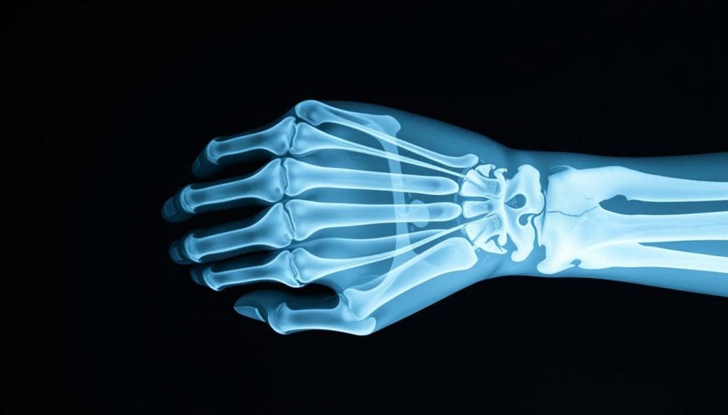
X-ray technology is key in diagnosing bone fractures. It uses electromagnetic waves to show the body’s inside, focusing on bones.
How X-Rays Work to Visualize Bone Structures
X-rays send radiation through the body. Denser materials like bone absorb more, showing up white. Softer tissues let more X-rays through, appearing gray or black. This helps doctors spot fractures and bone issues.
Strengths of X-Ray Imaging in Fracture Diagnosis
X-ray imaging is quick and widely available, perfect for emergencies. It clearly shows bone structures, helping find most fractures. Plus, it’s cheaper than other imaging like MRI.
Limitations and Blind Spots of X-Ray Technology
Despite its benefits, X-rays have limits. They might miss hairline or stress fractures. Also, they don’t show soft tissues well, which can be a problem for assessing injuries. For complex cases or soft tissue checks, doctors might suggest MRI. If you’re unsure about your imaging needs, talk to a healthcare expert.
The Science Behind MRI Bone Imaging
MRI technology is key in orthopedic diagnostics, giving deep insights into bone health. It works by creating detailed images of bones and soft tissues.
Magnetic Resonance Imaging Explained
MRI uses a strong magnetic field and radio waves to show body structures inside. It’s safer than X-rays because it doesn’t use harmful radiation. This is great for patients who need to be checked often.
Key components of MRI technology include:
- A powerful magnet that aligns the hydrogen atoms in the body
- Radiofrequency coils that transmit and receive signals
- A computer system that reconstructs the images
How MRI Visualizes Bone and Surrounding Tissues
MRI can see both bones and soft tissues, making it a top tool for doctors. It spots changes in bone marrow, cartilage, and ligaments. For example, it can find bone marrow edema, which means fluid in the bone marrow and might mean a fracture.
With MRI’s clear images, doctors can better understand injuries. This is key when an X-ray doesn’t show the full picture.
Types of MRI Sequences Used for Bone Imaging
There are different MRI sequences for looking at bones and soft tissues. Some common ones are:
- T1-weighted images, which show detailed anatomy
- T2-weighted images, which show inflammation or edema
- STIR (Short-Tau Inversion Recovery) sequences, which help find bone marrow lesions
Choosing the right MRI sequence helps doctors understand injuries better. This is vital for making the right treatment plan.
As we learn more about MRI’s role in finding broken bones, it’s clear it’s better than X-rays for many cases. This is true for complex injuries or when soft tissue damage is suspected.
Do MRI Show Broken Bones? Capabilities and Evidence
The role of MRI in finding bone fractures is a big topic in medicine. As we get better at medical imaging, knowing what MRI can and can’t do is key.
Detection Capabilities for Different Types of Fractures
MRI is great at finding many kinds of fractures, even those X-rays miss. It’s good at spotting fractures in soft tissues and bone marrow, which helps find hidden fractures.
Stress and hairline fractures are hard to see on X-rays early on. But MRI can spot them because it sees changes in bone marrow. A study on PubMed Central shows MRI’s power in finding these fractures.
Research on MRI Sensitivity and Specificity for Bone Fractures
Many studies have looked at MRI’s ability to find fractures. It’s been found to be 100% sensitive for some fractures, with accuracy rates of 95% to 100% in some areas.
Its high sensitivity comes from spotting changes in bone marrow early. This is really helpful for tricky areas like the spine and pelvis.
Clinical Studies Supporting MRI’s 95-100% Accuracy Rate
Many clinical studies back up MRI’s accuracy in finding fractures. They show MRI can spot fractures in hard-to-see areas, where X-rays fall short.
- MRI is very accurate in finding wrist and scaphoid bone fractures, which X-rays often miss.
- It’s also good at finding hip fractures in older patients.
- For athletes, MRI helps catch stress fractures early, allowing for quick treatment.
Using MRI helps doctors make better diagnoses. This leads to quicker and more effective treatment for fracture patients.
What Does an MRI Show That an X-Ray Cannot?
MRI technology can spot fractures and injuries that X-rays miss. This makes MRI key in diagnosing orthopedic and sports medicine issues.
Detecting Hairline and Stress Fractures
Hairline and stress fractures are hard to see with X-rays. MRI can spot these tiny fractures because it sees changes in bone marrow.
MRI is great for finding stress fractures in athletes or those with repetitive injuries. It shows bone marrow edema, helping diagnose and treat early.
Visualizing Bone Marrow Edema and Microfractures
Bone marrow edema is inflammation in the bone. MRI can see this condition that X-rays can’t.
Seeing bone marrow edema is key for treating bone injuries. It lets doctors start treatment early to prevent further damage.
Identifying Associated Soft Tissue Injuries
MRI can see soft tissues like ligaments, tendons, and muscles. This is important for finding soft tissue injuries with fractures.
MRI gives a full view of bone and soft tissue injuries. This helps create better treatment plans, even for complex cases.
Clinical Applications: When Will an MRI Show a Fracture Better Than X-Ray?
MRI technology is very useful in diagnosing fractures in certain situations. We will look at when MRI is better than X-ray. This helps in getting a more accurate diagnosis and better care for patients.
Specific Anatomical Regions Where MRI Excels
MRI is great for seeing fractures in tricky areas. For example, in the wrist and scaphoid area, MRI spots small fractures X-ray can’t. It’s also key for the hip and pelvis, showing both bones and soft tissues. This is vital for finding hidden fractures and understanding how bad the injury is.
- Detection of occult fractures in the hip and pelvis
- Visualization of scaphoid fractures in the wrist
- Assessment of fractures in the spine, including vertebral body and posterior element fractures
Sports Medicine Applications
In sports medicine, MRI is key for diagnosing and managing fractures. Stress fractures are hard to see on X-ray but MRI finds them easily. It also helps figure out how serious the fracture is and what treatment is best.
- Early detection of stress fractures
- Assessment of associated soft tissue injuries
- Monitoring of fracture healing and guiding return-to-play decisions
Pediatric Fracture Assessment
In kids, MRI is used more because it’s safer than X-ray. It’s great for finding fractures that X-ray can’t see. This is very important for checking non-accidental injuries in children.
Knowing when to use MRI over X-ray helps doctors make better choices. This leads to better care for patients. MRI is very useful in many areas, from specific body parts to sports medicine and kids’ health.
Practical Considerations: X-Ray vs. MRI for Fracture Diagnosis
Choosing between X-ray and MRI for fracture diagnosis has many practical aspects. Healthcare providers must consider the pros and cons of each to make the best choice.
Time and Cost Factors
Time and cost are key when deciding between X-ray and MRI. X-rays are quicker and cheaper than MRI scans. They are often the first choice for many cases. But, MRI offers detailed info that can be very helpful in some cases.
While MRI costs more, it can lead to more accurate diagnoses. This can save money in the long run.
Accessibility and Availability
X-ray machines are common in many medical places. They are found in emergency departments and primary care offices. MRI machines, though, are less common and need a special visit.
This makes X-rays a better choice for urgent cases where quick diagnosis is needed.
Patient Experience Differences
X-rays are quick and easy for patients. MRI scans, though, can be tough because of the machine’s size and the need to stay very quiet for a long time.
Patients with claustrophobia or anxiety may find MRI scans very hard. They might need extra help or sedation to finish the scan.
Radiation Exposure Comparison
MRI is safer than X-ray because it doesn’t use ionizing radiation. MRI uses a strong magnetic field and radio waves to create images. This makes it safer for people who need many scans or are sensitive to radiation, like children and pregnant women.
X-rays, on the other hand, do involve some radiation. While it’s usually safe, it can be a worry for some people over time.
Clinical Decision-Making: When to Use Each Imaging Modality
Clinicians must consider the pros and cons of X-ray and MRI for diagnosing fractures. The choice between them depends on the injury type, patient symptoms, and the situation. It’s all about making the right call for each patient.
First-Line Diagnostic Approaches
X-rays are usually the first choice for checking bone fractures. They’re fast, easy to find, and show bone health well. X-rays work great for checking bones in arms and legs. A study in the Journal of Orthopaedic Trauma showed X-rays are good at finding fractures in the wrist.
- Rapid assessment
- Wide availability
- Effective for initial fracture detection
When to Escalate from X-Ray to MRI
But sometimes, moving to MRI is needed. If pain persists after a negative X-ray, or if soft tissue injuries are suspected, MRI is better. MRI can spot bone marrow edema, stress fractures, and soft tissue injuries that X-rays miss.
For instance, MRI is key when hip pain doesn’t show up on X-rays. A study in the American Journal of Roentgenology showed MRI can find hip fractures X-rays can’t.
Emergency vs. Non-Emergency Scenarios
In emergency cases, like sudden trauma, X-rays are often first because they’re quick and easy. But in non-emergency cases, where there’s time to think, MRI might be chosen based on symptoms and X-ray results.
“The choice of imaging modality should be guided by the clinical context and the need for detailed soft tissue evaluation.”Orthopedic Surgeon
Insurance and Cost-Effectiveness Considerations
Insurance and cost also play a big role. MRI gives more detailed info but costs more than X-ray. Insurance policies vary, affecting the choice. Studies often suggest starting with X-ray and using MRI only when needed.
By thinking about these points, doctors can decide wisely between X-ray and MRI. This helps improve patient care.
Conclusion: The Complementary Role of X-Ray and MRI in Fracture Diagnosis
We’ve looked at how X-ray and MRI help find bone fractures. We’ve seen their good points and what they can’t do. Now, we know MRI is great at showing fractures, even when X-rays can’t.
When thinking about “can an MRI show broken bones,” the answer is yes. It’s very good at spotting hairline and stress fractures. Plus, it sees soft tissue injuries well. This makes MRI a key tool for doctors.
Knowing “what does an MRI show that an X-ray can’t” helps doctors pick the best test. MRI is top-notch at finding bone marrow edema and tiny fractures. It gives a full view of how bad a fracture is and any other injuries.
In short, X-ray and MRI work together to diagnose fractures. By picking the right test for each case, doctors can give the best care. This leads to better health for patients.
FAQ
What does an MRI show that an X-ray can’t?
MRI can spot small fractures, bone marrow swelling, and soft tissue injuries. These might not show up on an X-ray.
Can MRI detect broken bones?
Yes, MRI is great at finding different kinds of fractures. This includes tiny and stress fractures.
Will an MRI show a broken bone?
Yes, MRI can reveal broken bones. It often shows more detail than X-rays, even when fractures are hard to see.
Do MRI show broken bones?
Yes, MRI can show broken bones. It’s very useful for finding fractures that X-rays miss.
Does MRI show bone fractures?
Yes, MRI is very good at finding bone fractures. It can spot fractures that X-rays can’t see.
What are the advantages of MRI over X-ray in detecting fractures?
MRI has several benefits. It can find hairline fractures, see bone marrow swelling, and spot soft tissue injuries.
When is MRI preferred over X-ray for fracture detection?
MRI is often chosen for certain areas, sports medicine, and kids. It’s needed for detailed images.
How does MRI compare to X-ray in terms of radiation exposure?
MRI doesn’t use radiation. This makes it safer for patients, even when they need more scans.
Can an MRI show a fracture that an X-ray misses?
Yes, MRI can find fractures that X-rays can’t. This is true for small or hidden fractures.
What factors influence the choice between X-ray and MRI for fracture diagnosis?
The choice between X-ray and MRI depends on several things. These include time, cost, and how easy it is to get the scan. Patient experience and the situation also matter.
Reference
- Ghosh, S., et al. (2024). Automated bone fracture detection in X-ray imaging to improve diagnostic efficiency. Journal of Digital Imaging. https://www.sciencedirect.com/science/article/pii/S187705092400632X


