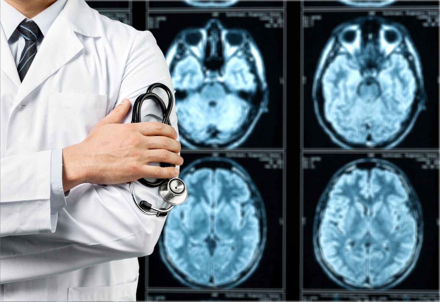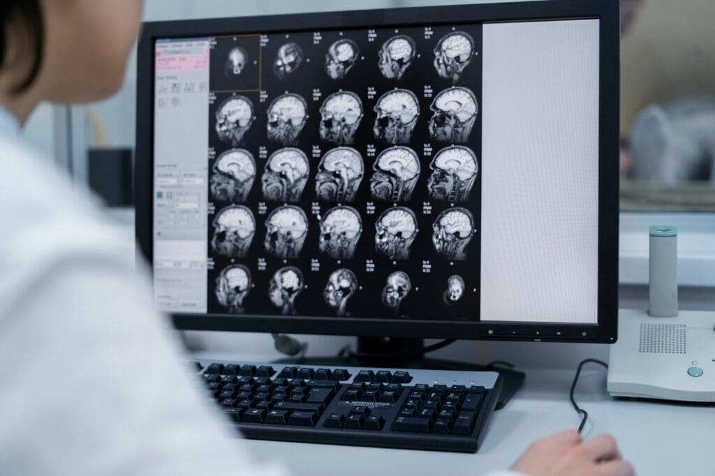
When there’s worry about a brain hemorrhage, a quick CT scan can save lives. It gives clear results to help start treatment right away. At Liv Hospital, we know how important CT scans are in emergencies. We’re dedicated to helping international patients get top-notch medical care.
A CT scan is key for spotting intracranial hemorrhage fast. This includes bleeding in the brain caused by injury or stroke. Our advanced neuroimaging services help us give our patients the best care possible.
Key Takeaways
- Rapid CT scans are crucial for detecting brain bleeds in emergency situations.
- CT scans can identify various types of intracranial hemorrhages.
- Accurate diagnosis through CT scans guides effective treatment plans.
- Liv Hospital is committed to providing world-class neuroimaging services.
- Our expertise supports international patients seeking advanced medical care.
What Does a CT Scan of the Brain Show?

CT scans of the brain are key in emergency care. They quickly and accurately check for brain injuries and conditions. This imaging helps us diagnose and treat neurological emergencies fast.
Basic Principles of Brain CT Imaging
Brain CT imaging uses X-rays to make detailed images of the brain. It shows the brain’s parts and any problems like hemorrhages or tumors.
CT scans work by seeing how X-rays are absorbed by different tissues. This lets them show different brain tissues and problems. It gives us a clear view of what’s happening in the brain.
Why CT Scans Are Preferred for Emergency Brain Assessment
CT scans are fast and available, making them great for emergencies. They’re especially good at finding acute hemorrhages and other urgent issues. This makes them essential for checking patients with brain injuries or conditions.
One big plus of CT scans is how quickly they can image the brain. This helps us diagnose and treat fast, especially in traumatic brain injuries or strokes. Quick action can greatly improve outcomes.
| Advantages of CT Scans | Description |
|---|---|
| Speed | Quick imaging process, crucial in emergencies |
| Sensitivity | Highly sensitive to acute hemorrhages and other emergencies |
| Availability | Widely available in most medical facilities |
How Quickly Results Are Available in Urgent Situations
In urgent cases, CT scan results come back in minutes. This is because the images are quickly taken and can be read right away by doctors.
This fast result time lets doctors make quick decisions. They can decide on surgery, more tests, or other treatments right away.
Understanding what a CT scan of the brain shows is key. It helps us diagnose and manage many neurological conditions. From bleeding to structural issues, CT scans give us the info we need to make treatment plans.
Acute Intracranial Hemorrhage Detection

CT scans have changed emergency medicine by quickly spotting intracranial hemorrhage. They give us fast and accurate diagnoses in emergencies.
Subarachnoid Hemorrhage Appearance on CT
Subarachnoid hemorrhage means bleeding around the brain. On a CT scan, it shows up as bright blood in the brain’s spaces. We look for this to spot subarachnoid hemorrhage.
The look of subarachnoid hemorrhage on CT scans changes with blood amount and time. In the early stages, the blood is usually brighter than the brain.
Intracerebral Hemorrhage Characteristics
Intracerebral hemorrhage is blood inside the brain. On CT scans, it looks like a bright spot with clear edges. We check its size, where it is, and any pressure it might cause.
| Characteristic | Description |
|---|---|
| Appearance | Hyperdense lesion on CT |
| Location | Within brain parenchyma |
| Margins | Well-defined |
Subdural and Epidural Hematoma Identification
Subdural hematomas are blood between the dura mater and arachnoid mater. Epidural hematomas are between the dura mater and the skull. On CT scans, subdural hematomas look like crescents, and epidural hematomas are biconvex.
We study CT scans closely to tell subdural and epidural hematomas apart. Their treatment can be very different.
Traumatic Brain Injuries Visible on CT Scans
CT scans have changed how we diagnose traumatic brain injuries. They offer a fast and effective way to check injury severity. When someone has a head injury, a CT scan is often the first step to see how bad the damage is.
Major Skull Fractures and Associated Bleeding
Identifying major skull fractures is key in assessing traumatic brain injuries. These fractures can lead to significant bleeding, either in the brain or around it. CT scans are great at finding these fractures and the bleeding they cause, helping doctors act quickly.
“Being able to quickly and accurately spot skull fractures and bleeding is vital in treating traumatic brain injuries,” says a top neurosurgeon. “CT scans give the details needed to make treatment decisions.”
Contusions and Impact Injuries
Contusions, or brain bruises, are common in traumatic brain injuries. They happen when the brain hits the skull, damaging brain tissue. CT scans can show these contusions and other impact injuries, helping doctors understand the injury’s full extent.
Subtle Hemorrhages That May Indicate Trauma
CT scans can also find small hemorrhages that might show trauma. These small bleeds can be an early warning of bigger problems. Modern CT scanners can spot even small hemorrhages, making sure patients get the right care.
CT scans give a detailed look at the brain and find both big and small injuries. This is crucial for creating a good treatment plan and improving patient outcomes.
CT Scan Brain Bleed Findings After Stroke
CT scans are key in diagnosing strokes, giving quick insights into brain bleed findings. Timely and accurate diagnosis is vital for effective treatment.
Hemorrhagic Stroke Visualization
Hemorrhagic strokes are caused by bleeding in or around the brain. On a CT scan, this shows up as bright spots from fresh blood. “The ability of CT scans to quickly identify hemorrhagic strokes is unparalleled,” says a leading neurologist. “This rapid diagnosis is critical for guiding immediate treatment decisions.”
The location and size of the hemorrhage are crucial. They help determine the stroke’s severity and the best treatment. CT scans can accurately depict the extent of bleeding, helping clinicians to assess the situation effectively.
Ischemic Stroke Patterns and Evolution
Ischemic strokes happen when an artery to the brain gets blocked. Early CT scans might look normal or show small signs of ischemia. But as the stroke area grows, changes become clearer on later scans.
Early detection of ischemic changes is vital for starting the right therapy, like thrombolysis, to reduce brain damage. Our experts stress the need for serial CT scans to track ischemic strokes.
Differentiating New vs. Old Stroke Changes
Distinguishing new from old stroke changes on a CT scan is a challenge. New strokes show up as areas of acute hemorrhage or ischemia. Older strokes might look like softening of the brain tissue or scarring.
“The differentiation between acute and chronic stroke changes on CT scans is crucial for clinical decision-making,” notes a radiologist with extensive experience in neuroradiology.
Healthcare professionals analyze CT scan findings to figure out the stroke’s timing and nature. This helps guide treatment and rehabilitation plans.
What Can a CT Scan Show in the Brain: Space-Occupying Lesions
Space-occupying lesions in the brain can be very dangerous. CT scans are often the first step to find them. These lesions can be tumors, vascular malformations, or cysts. Each type shows different things on a CT scan.
Brain Tumor Detection and Characteristics
CT scans can find brain tumors, whether they are benign or malignant. We look at the tumor’s location, size, and density. Some tumors might show up more clearly with contrast, meaning they have more blood vessels.
Brain tumors can look different on CT scans. For example, meningiomas are usually dense and might have calcium spots. Gliomas, on the other hand, can look like mixed-up masses with dead areas.
| Tumor Type | Typical CT Appearance | Contrast Enhancement |
|---|---|---|
| Meningioma | Dense, well-defined | Often strong |
| Gliomas | Heterogeneous, ill-defined | Variable |
| Metastasis | Multiple, variable density | Often ring-enhancing |
Arteriovenous Malformations and Aneurysms
Arteriovenous malformations (AVMs) and aneurysms can be seen on CT scans, especially with contrast. AVMs look like a mess of blood vessels, sometimes with calcium spots. Aneurysms are seen as swellings in arteries.
CT angiography helps us see these vascular problems better. This is important for planning treatment.
Cysts and Other Non-Hemorrhagic Masses
CT scans can also find cysts and other non-hemorrhagic masses. These include arachnoid cysts, colloid cysts, and epidermoid cysts. Each type looks different on a CT scan.
Arachnoid cysts are usually the same density as CSF and push against brain structures. Epidermoid cysts might look more mixed up because of their contents.
By spotting these lesions, we can help plan better treatment. This can lead to better outcomes for patients.
Signs of Elevated Intracranial Pressure
Elevated intracranial pressure shows up in different ways on a CT scan. These signs are key for catching the problem early and stopping more brain damage.
Ventricular System Compression or Enlargement
The ventricular system is very sensitive to changes in pressure. Compression or enlargement of the ventricles means there’s high pressure. We see these changes on CT scans to check the ventricular system’s health.
High pressure can squeeze or swell the ventricles. This happens when cerebrospinal fluid (CSF) pathways get blocked. A CT scan can show this, helping us diagnose.
Midline Shift Measurement and Significance
Midline shift is a big warning sign of high pressure. It happens when a mass effect pushes brain structures across the midline. Measuring the extent of midline shift helps us understand how serious it is.
CT scans measure midline shift by looking at the displacement of the septum pellucidum or the pineal gland. A shift over 5 mm is serious and might need quick action.
| Midline Shift Measurement | Clinical Significance |
|---|---|
| 0-5 mm | Mild, usually monitored |
| 5-10 mm | Moderate, may require intervention |
| >10 mm | Severe, often requires urgent intervention |
Brain Edema Patterns and Severity Assessment
Brain edema, or swelling, often comes with high pressure. Assessing the patterns and severity of brain edema is crucial for knowing the extent of injury.
We check brain edema on CT scans by spotting hypodensity areas. The severity depends on how big and where the edema is, along with any mass effect or midline shift.
Knowing these signs helps us diagnose and manage high intracranial pressure well.
What Will a CT Scan of the Brain Show: Anatomical Abnormalities
A CT scan of the brain is a key tool for finding different problems. It gives us detailed views of the brain’s structure. This helps us diagnose and treat many conditions.
These problems can come from being born with them, getting sick, or after surgery. Knowing about these issues is key for good patient care.
Congenital Malformations
Congenital malformations are problems present at birth. CT scans can spot these, like hydrocephalus, Chiari malformations, or arachnoid cysts. Finding them early lets doctors plan the right treatment.
Hydrocephalus Detection and Monitoring
Hydrocephalus is when too much cerebrospinal fluid builds up in the brain. This causes high pressure. CT scans help find this by showing big ventricles.
They also help check if treatments, like shunts, are working. Seeing how ventricles change over time is key for managing hydrocephalus. Regular scans help adjust treatment plans for the best results.
Post-Surgical Anatomical Changes
After brain surgery, CT scans check for changes. These can include surgical tools, changes from tumor removal, or complications like bleeding or swelling.
CT scans watch the healing and spot problems early. This info is crucial for post-surgery care. It helps patients recover as well as possible.
What Can a Head CT Scan Show: Post-Treatment Monitoring
We use head CT scans to keep a close eye on patients after treatment. This is key to making sure they get the best care. It lets us see how well the treatment is working and spot any problems early.
CT scans give us important information about the brain’s state after treatment. This helps us decide on the next steps in care.
Post-Operative Hemorrhage Detection
CT scans are vital for finding bleeding in the brain after surgery. Bleeding can be very dangerous and needs quick action. They help us spot bleeding by looking for dense areas on the scan.
Evaluating Effectiveness of Interventions
CT scans help us see if treatments are working. We compare scans before and after treatment. For example, they show if a tumor has shrunk, meaning the treatment was successful.
We use these scans to fine-tune treatment plans. This ensures the best results for the patient.
Identifying Treatment Complications
CT scans also help find problems caused by treatment. Issues like infection or swelling can happen. By watching for these, we can act fast to help.
The table below shows some common problems and how they look on CT scans.
| Complication | CT Scan Appearance |
|---|---|
| Infection | Hypodense areas with possible enhancement |
| Edema | Hypodense areas around the treated region |
| Hemorrhage | Hyperdense areas |
Limitations of CT Scans in Brain Bleed Assessment
CT scans are very useful but have some limits. They are key in emergency brain care. Knowing these limits is important for doctors and patients.
When MRI May Be Preferred Over CT
MRI is better than CT scans in some ways. It’s more sensitive to brain changes and can spot small hemorrhages. MRI is also better for the brainstem and areas CT scans can’t see well.
MRI vs. CT: The choice between MRI and CT depends on the situation. MRI is better for detailed images, like in chronic cases. But, MRI takes longer and isn’t always available.
Small Hemorrhages That May Be Missed
CT scans might miss small hemorrhages. These can be from various causes, like small vessel disease. The small size and location of these bleeds can make them hard to see on CT scans.
Clinical Implication: Not seeing small hemorrhages can be serious. It might mean there’s a problem that needs attention. A follow-up MRI might be needed to check for these small bleeds.
Does a CT Scan Show Brain Activity?
CT scans are for structural issues, not brain activity. They can show structural problems or hemorrhages but not how the brain works.
For brain activity, other tests like fMRI or EEG are better. They show how the brain functions, adding to what CT scans reveal.
Understanding neuroimaging can be tough. Knowing CT scan limits and when to use other tests helps doctors make better choices. This leads to better care for patients.
Conclusion: The Critical Role of CT Scans in Brain Hemorrhage Diagnosis
CT scans are key in finding brain hemorrhages. They can spot acute intracranial hemorrhage, traumatic brain injuries, and stroke quickly and accurately. We offer top-notch healthcare to patients from around the world.
CT scans help doctors make fast, smart choices about patient care. They give us the details we need to create treatment plans that fit each patient’s needs. This approach helps improve care and save lives.
In short, CT scans are vital for diagnosing and treating brain hemorrhages. We keep using this technology to give our patients the best care. We aim to ensure they get the best treatment and support every step of the way.
FAQ
What does a brain CT scan show?
A brain CT scan gives detailed images of the brain. It can spot abnormalities like bleeds, tumors, or injuries. This tool helps us quickly check the brain’s condition in emergencies.
What can a CT scan show in the brain?
A CT scan can reveal many conditions. It can spot hemorrhages, tumors, cysts, and signs of high pressure in the brain. Our team uses these images to make accurate diagnoses and treatment plans.
Can a CT scan detect brain bleeds?
Yes, a CT scan is great at finding brain bleeds. It can spot subarachnoid hemorrhage, intracerebral hemorrhage, and other types of bleeding. We count on CT scans to quickly find these serious conditions.
What is the difference between a CT scan and an MRI for brain assessment?
Both CT scans and MRIs are useful, but CT scans are faster. They’re better for emergency situations because they can quickly find acute hemorrhages. MRI might be used for other conditions or for more detailed checks.
Can a CT scan show brain activity?
No, a CT scan doesn’t show brain activity. It gives images of the brain’s structure but doesn’t check how it’s working. Other tests, like EEG or functional MRI, are needed to see brain activity.
How quickly are CT scan results available?
CT scan results are usually ready fast, often in minutes. This is key in emergencies where quick diagnosis and treatment are crucial.
What are the limitations of CT scans in brain bleed assessment?
CT scans are very good, but they might miss small hemorrhages or certain injuries. Sometimes, MRI is used to get more info or to check the brain further.
Can a CT scan detect traumatic brain injuries?
Yes, a CT scan can find many types of traumatic brain injuries. It can spot skull fractures, contusions, and hemorrhages. Our team uses CT scans to see how severe these injuries are and plan treatment.
What can a head CT scan show in terms of post-treatment monitoring?
A head CT scan can check on how a patient is doing after treatment. It can spot post-operative bleeding, see if treatments are working, and find any complications. We use CT scans to keep an eye on patients and adjust treatment as needed.
Can a CT scan detect anatomical abnormalities?
Yes, a CT scan can find many anatomical issues. It can spot congenital malformations, hydrocephalus, and changes after surgery. Our experts look at CT scan images to find these problems and plan treatment.
What does a CT scan of the head show?
A CT scan of the head shows detailed images of the brain and its surroundings. It can find abnormalities or injuries. We use it to check for a range of conditions, from brain injuries to tumors.
References
- Surgery for Intracerebral Hemorrhage. Retrieved from: https://neurosurgery.weillcornell.org/condition/intracerebral-hemorrhage/surgery-intracerebral-hemorrhage
- Intracerebral Hemorrhage. Retrieved from: https://www.upmc.com/services/neurosurgery/brain/conditions/neurovascular-conditions/conditions/intracerebral-hemorrhage
- Traumatic Brain Injuries. Retrieved from: https://www.cdc.gov/traumatic-brain-injury/index.html


