A cerebral arteriovenous malformation (AVM) is a tangled mess of blood vessels in the brain. Arteries connect directly to veins, skipping capillaries. This setup creates high pressure, leading to serious problems.
Usually, arteries bring oxygen-rich blood to the brain. Veins take oxygen-poor blood back to the heart and lungs. But a cerebral AVM messes with this, raising the chance of a rupture and severe effects.
At Liv Hospital, we know the dangers of a ruptured brain AVM. We’re dedicated to top-notch care and the latest treatments for this condition.
Key Takeaways
- A cerebral AVM is an abnormal connection between arteries and veins in the brain.
- This condition can lead to a high-pressure system, increasing the risk of rupture.
- A ruptured brain AVM can have severe and sudden consequences.
- Early recognition and expert care are critical in managing this condition.
- Liv Hospital offers advanced treatment options for cerebral AVMs with a patient-centered approach.
Understanding Cerebral Arteriovenous Malformations

Cerebral arteriovenous malformations (AVMs) are abnormal connections between arteries and veins in the brain. They disrupt normal blood flow and can lead to neurological issues. It’s important to understand these vascular anomalies to diagnose and treat them well.
Definition and Basic Structure
A cerebral arteriovenous malformation is a tangle of blood vessels in the brain. It connects arteries directly to veins, skipping the capillary system. This can cause high-pressure blood flow and a risk of rupture.
AVMs can happen anywhere in the brain, including the cerebellum. They are called cerebellar arteriovenous malformations there. An AVM has feeding arteries and draining veins.
How Normal Brain Blood Flow Works
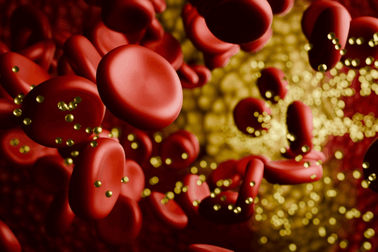
Normal brain blood flow is carefully regulated. It ensures the brain gets the oxygen and nutrients it needs. Arteries bring oxygenated blood to the brain, where it goes through capillaries for exchange. Then, deoxygenated blood is carried away by veins.
How AVMs Disrupt Normal Blood Flow
AVMs disrupt normal blood flow. The direct connection between arteries and veins means blood skips capillaries. This can lead to reduced oxygen delivery to brain tissues. It causes neurological symptoms and increases the risk of hemorrhage.
| Aspect | Normal Brain Blood Flow | Blood Flow with AVM |
|---|---|---|
| Oxygen Delivery | Optimal oxygen delivery through capillaries | Reduced oxygen delivery due to bypassing capillaries |
| Blood Pressure | Regulated blood pressure | High-pressure blood flow through AVM |
| Risk of Hemorrhage | Low risk | Increased risk due to high pressure and structural weaknesses |
Understanding these differences is key to managing and treating cerebral AVMs effectively.
The Prevalence and Demographics of Cerebral AVMs
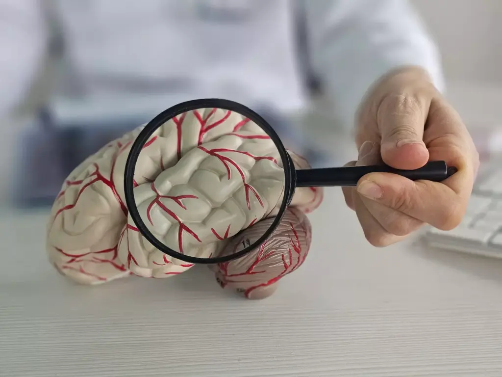
Cerebral AVMs are rare and can affect health a lot. Knowing how common they are helps us understand the risks and how to manage them.
Incidence Rates in the General Population
Less than 1% of people have cerebral AVMs. Research shows about 1-2 cases per 100,000 people each year. This rarity makes studying them hard.
Key statistics on cerebral AVM incidence include:
- Approximately 1 in 7000 people in the United States has a cerebral AVM.
- Cerebral AVMs account for about 2% of all hemorrhagic strokes.
- The condition is often congenital, meaning it is present at birth.
Age Groups Most Commonly Affected
Cerebral AVMs can happen to anyone, but they’re most common in young adults. They’re often found in people between their 20s and 40s, during tests or after a bleed.
Younger people are more likely to show symptoms like bleeding or seizures. Older adults usually show symptoms earlier in life.
Genetic and Congenital Factors
Most cerebral AVMs happen by chance, but genetics play a role. People with hereditary hemorrhagic telangiectasia (HHT) are more at risk. HHT affects blood vessel formation in the body, including the brain.
Genetic considerations include:
- Increased risk of cerebral AVMs in individuals with HHT.
- Potential for multiple AVMs in individuals with certain genetic conditions.
- Ongoing research into genetic markers that may predict the risk of AVM formation or rupture.
Types and Locations of Cerebral AVMs
It’s important to know where cerebral AVMs are found to treat them well. These malformations can be in different parts of the brain. Each area has its own challenges and risks.
Cerebellar Arteriovenous Malformations
Cerebellar AVMs are in the cerebellum. This part of the brain helps with balance and coordination. These AVMs can be tricky because of their location and the cerebellum’s important role.
Symptoms and Risks: People with cerebellar AVMs might feel dizzy, lose balance, or have trouble speaking. A big worry is that these AVMs could burst. This could cause serious bleeding and be very dangerous.
Cerebral Venous Malformations
Cerebral venous malformations (CVMs) affect the brain’s veins. They are usually not serious but can be linked to other problems. They might also bleed, which is a concern.
Diagnostic Challenges: Finding CVMs can be hard because they don’t always show symptoms. They might only be found when someone gets an imaging test for another reason.
Other Common Locations in the Brain
AVMs can also be in other brain areas like the cerebral hemispheres, basal ganglia, and thalamus. Each place has its own treatment challenges.
The table below shows where cerebral AVMs are found and what they might affect:
| Location | Characteristics | Risks |
|---|---|---|
| Cerebellum | Affects coordination and balance | High risk of rupture, severe hemorrhage |
| Cerebral Hemispheres | Can affect motor and sensory functions | Variable risk depending on size and location |
| Basal Ganglia/Thalamus | Involves critical brain structures | High risk due to deep location |
Knowing where and what a cerebral AVM is helps decide the best treatment. We’ll look at more about cerebral AVMs in the next parts.
The Anatomy of a Cerebral AVM
A cerebral AVM is an abnormal connection between arteries and veins in the brain. It lacks the normal capillary bed. This creates a complex vascular structure that can affect brain function and health.
Structural Abnormalities
The arteries and veins in an AVM are tangled, without the usual smaller blood vessels and capillaries. Blood flows directly from arteries to veins, making it a high-flow, low-resistance shunt.
This abnormal flow can cause several structural issues. These include:
- Enlarged feeding arteries due to increased blood flow
- Dilated draining veins from high pressure
- A nidus, the central part of the AVM with abnormal vessels
Size Variations and Classifications
Cerebral AVMs vary in size, from small to large. The size affects the risk of rupture and treatment complexity.
| Size Category | Description | Typical Characteristics |
|---|---|---|
| Small | Less than 3 cm in diameter | Often considered low risk, may be asymptomatic |
| Medium | 3-6 cm in diameter | Moderate risk, may cause symptoms due to mass effect or bleeding |
| Large | Greater than 6 cm in diameter | High risk, often symptomatic, and challenging to treat |
Feeding Arteries and Draining Veins
The feeding arteries supply blood to the AVM, while the draining veins carry it away. Knowing these vessels is key for treatment planning.
Feeding Arteries: These arteries are enlarged and tortuous, supplying a lot of blood to the AVM. Identifying and understanding these arteries is vital for treatments like embolization.
Draining Veins: The draining veins are dilated and under high pressure. Their anatomy affects the risk of rupture and treatment choice.
Risk Factors for Cerebral AVM Rupture
We need to look at the different risk factors for cerebral AVM rupture. This is to help those affected get better care. A ruptured AVM in the brain can lead to serious bleeding inside the skull. This bleeding can be very dangerous and may cause death or disability.
The chance of a brain AVM bleeding is about 2% to 3% each year. But, some types of AVMs might have a higher risk.
Previous Bleeding Episodes
A history of bleeding is a big risk factor for AVM rupture. Research shows that AVMs that have bled before are more likely to bleed again. This is something we must think about when managing cerebral AVMs.
Size and Location Considerations
The size and where an AVM is located also matter a lot. Bigger AVMs and those in deeper parts of the brain might bleed more easily. We’ll see how these factors affect the risk of rupture.
Deep Venous Drainage
Deep venous drainage can also raise the risk of AVM rupture. AVMs with this type of drainage are more likely to bleed. This is because the pressure inside the malformation is higher.
Associated Aneurysms
Having aneurysms with cerebral AVMs also increases the risk of rupture. Aneurysms are abnormal blood vessel dilations. They can be inside or near the AVM, making it more complicated and raising the risk of bleeding.
| Risk Factor | Description | Impact on Rupture Risk |
|---|---|---|
| Previous Bleeding Episodes | History of previous AVM bleeding | Increased risk of re-bleeding |
| Size and Location | Larger AVMs and deeper locations | Higher risk for larger and deeper AVMs |
| Deep Venous Drainage | Presence of deep venous drainage | Increased pressure, higher risk |
| Associated Aneurysms | Presence of aneurysms within or near AVM | Complicates AVM structure, increases risk |
Understanding the Annual Rupture Risk
The risk of AVM rupture is a big worry for both patients and doctors. This risk helps decide how to manage AVMs in patients.
Statistical Analysis of Rupture Rates
Research shows that unruptured AVMs have a 2-4% chance of bleeding each year. If an AVM has already ruptured, the risk jumps to about 17% annually. These numbers highlight the need to know each patient’s specific risk factors.
Researchers have done a lot of work to understand this risk. They look at things like the AVM’s size and location, and the patient’s health history.
| AVM Status | Annual Rupture Risk |
|---|---|
| Unruptured AVM | 2-4% |
| Previously Ruptured AVM | 17% |
Cumulative Lifetime Risk
It’s important to think about the lifetime risk of AVM rupture. This means looking at the chance of rupture over a patient’s whole life, considering their age and how long they might live.
Cumulative lifetime risk is a key factor in deciding treatment. Younger patients face a higher risk over their lifetime compared to older ones.
Risk Assessment Tools
There are tools to help doctors figure out the risk of AVM rupture. These tools look at things like the AVM’s size, where it is, and how it drains blood.
Using these tools, doctors can give patients advice and treatment plans that fit their unique situation.
What Happens During a Brain AVM Rupture
When a brain AVM ruptures, it’s because the blood vessel walls can’t handle the pressure. The high pressure makes the blood vessel walls thin and weak.
This pressure can cause the blood vessel to burst, leading to bleeding in or around the brain. This is a serious emergency that needs quick medical help.
The Mechanism of Bleeding
The bleeding in a brain AVM rupture happens when the weak blood vessel walls can’t take the pressure. When they burst, blood spills into the brain, causing damage and serious problems.
The bleeding can be categorized based on its location and severity. Knowing how it happens helps doctors find the best treatments.
Types of Intracranial Hemorrhage
Intracranial hemorrhage from a brain AVM rupture can be different based on where and how it bleeds.
| Type of Hemorrhage | Description |
|---|---|
| Intraparenchymal Hemorrhage | Bleeding into the brain tissue itself, which can lead to significant damage. |
| Subarachnoid Hemorrhage | Bleeding into the space surrounding the brain, which can cause severe headache and other symptoms. |
| Intraventricular Hemorrhage | Bleeding into the ventricles of the brain, which can lead to hydrocephalus. |
Immediate Physiological Effects
The effects of a brain AVM rupture are immediate and serious. The bleeding can increase pressure in the brain, damage brain tissue, and disrupt brain function.
Symptoms can range from sudden, severe headache to seizures, neurological deficits, and even loss of consciousness. Quick medical care is key to reducing these effects and improving chances of recovery.
Recognizing Symptoms of a Ruptured Brain AVM
A ruptured brain AVM can show severe symptoms that need quick action. It’s a life-threatening situation that requires immediate medical help.
Sudden, Severe Headache
One common symptom is a sudden, severe headache. People often say it’s “the worst headache of my life.” This headache is very intense and may come with nausea and vomiting.
Seizures and Neurological Deficits
Ruptured brain AVMs can cause seizures and neurological problems. These problems depend on where and how bad the bleed is. They might include weakness, numbness, speech or vision issues, and changes in thinking.
Some people with brain AVMs may have symptoms like seizures, headaches, or muscle weakness. These happen because of the abnormal blood flow and pressure in the AVM.
Loss of Consciousness
In severe cases, a ruptured brain AVM can cause loss of consciousness. This is a medical emergency that needs quick action to prevent more brain damage or death.
Warning Signs Before Rupture
Some people with brain AVMs may have warning signs before it ruptures. These can include headaches, seizures, or other neurological symptoms. It’s important for those with known AVMs to watch for these signs and seek medical help if they happen.
| Symptom | Description | Frequency |
|---|---|---|
| Sudden, Severe Headache | Often described as “the worst headache of my life” | Common |
| Seizures | Can occur due to abnormal electrical activity | Frequent |
| Neurological Deficits | Weakness, numbness, speech or vision difficulties | Variable |
| Loss of Consciousness | Medical emergency requiring immediate attention | Less Common |
Diagnosing Cerebral AVMs: Advanced Imaging Techniques
Diagnosing cerebral arteriovenous malformations (AVMs) needs advanced imaging and clinical checks. These imaging tools help find AVMs, their size, and where they are. This info is key for choosing the right treatment.
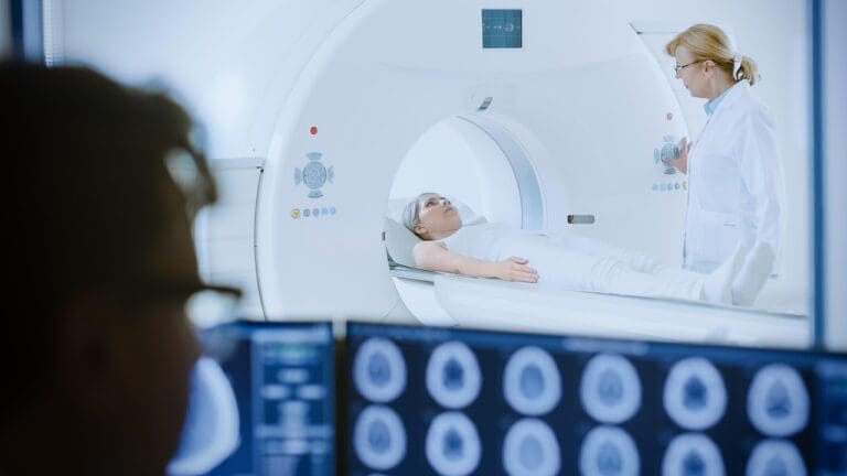
Magnetic Resonance Imaging (MRI)
MRI is a non-invasive way to see the brain’s soft tissues clearly. It’s great for spotting AVMs’ problems, like the nidus and blood flow issues.
Computed Tomography (CT)
CT scans help see AVMs quickly, which is important in emergencies. CT angiography gives detailed blood vessel images, helping spot AVMs.
Cerebral Angiography
Cerebral angiography is the top choice for AVM diagnosis. It involves putting contrast into blood vessels to see the AVM’s details.
Clinical Evaluation Process
The clinical check-up looks at the patient’s history, symptoms, and physical exam. This info, with imaging, helps doctors diagnose AVMs and plan treatment.
Treatment Options for Unruptured and Ruptured Cerebral AVMs
When it comes to treating cerebral AVMs, we have several options. These choices depend on the AVM’s size, location, and the patient’s health. We also look at the risk of rupture or re-rupture.
Surgical Resection
Surgical resection is a key treatment for cerebral AVMs. It involves removing the malformation surgically. This method is best for AVMs that are easily reached and at high risk of rupture.
Microsurgical techniques help us remove the AVM carefully. This way, we avoid harming the surrounding brain tissue.
Endovascular Embolization
Endovascular embolization is a less invasive option. It uses a catheter to block the AVM with embolic materials. This method can be used alone or with other treatments like surgery or radiosurgery.
Embolization can shrink the AVM. This makes it easier to treat with other methods.
Stereotactic Radiosurgery
Stereotactic radiosurgery is a non-surgical treatment. It uses focused radiation to destroy the AVM. This method is good for AVMs in hard-to-reach areas of the brain.
Radiosurgery aims to completely remove the AVM over time. It’s a safe and effective option for many patients.
Conservative Management Approaches
In some cases, conservative management is recommended. This is for patients with unruptured AVMs that are not causing symptoms. We monitor these AVMs with imaging studies.
Lifestyle modifications and managing related conditions, like high blood pressure, are also part of conservative management.
We work with each patient to find the best treatment plan. We consider their unique situation and needs. This way, we offer personalized care for those with cerebral AVMs.
Living with an Unruptured Cerebral AVM: Management Strategies
Living with an unruptured AVM can be tough, but the right strategies help. At Liv Hospital, we offer top-notch diagnosis and treatment for cerebral AVMs. We provide full care for our patients.
Monitoring Protocols
Choosing the right action for an unruptured AVM is hard. It’s found on tests but doesn’t cause symptoms. Regular checks are key. We use MRI or CT scans to watch for size or structure changes.
We suggest patients stay close to their doctors. This way, they can set up a monitoring plan that fits their needs.
Lifestyle Modifications
Changing your lifestyle can help manage risks from an unruptured AVM. Avoiding head trauma, like in contact sports, is important. Also, managing stress with relaxation techniques is helpful.
Key lifestyle changes include:
- Avoid smoking and drinking too much alcohol
- Eat healthy and exercise regularly
- Keep blood pressure and heart risks under control
Medication Management
There’s no special medicine for an unruptured AVM. But, managing related health issues is key. This includes controlling blood pressure and using seizure meds if needed.
We help our patients create a medication plan that meets their health needs.
Psychological Impact and Support
Dealing with an unruptured AVM can really affect your mind. It can cause anxiety and stress. It’s vital to have support, like counseling and support groups.
At Liv Hospital, we believe in caring for the whole person. We offer resources to support our patients’ mental and emotional health.
Conclusion
We’ve looked into cerebral arteriovenous malformations, a complex condition needing careful handling. It’s key for both patients and doctors to understand cerebral AVMs well. This knowledge helps in making smart choices about diagnosis, treatment, and care.
A summary of cerebral AVMs shows how vital it is to spot symptoms early. Knowing how to diagnose and the treatment options available is also important. The treatment plan varies based on the AVM’s size, location, and risk of bleeding.
Our talk has stressed the need for a detailed approach to managing cerebral AVMs. This approach considers the person’s health and their specific AVM situation. We aim to give patients the knowledge they need to handle their treatment journey well.
Managing cerebral AVMs effectively needs a team of healthcare experts working together. They provide care tailored to each patient. We think informed patients can better manage their condition and make treatment choices.
What is a cerebral arteriovenous malformation (AVM)?
A cerebral arteriovenous malformation (AVM) is a mix of blood vessels in the brain. It can disrupt blood flow, leading to complications like rupture and bleeding.
How common are cerebral AVMs?
Cerebral AVMs are rare, affecting about 1 in 100,000 people each year. They can be present at birth or develop later.
What are the risk factors for a cerebral AVM rupture?
Risk factors for a cerebral AVM rupture include previous bleeding, larger AVM size, and deep venous drainage. Knowing these factors helps in managing and treating AVMs.
What happens during a brain AVM rupture?
During a brain AVM rupture, bleeding occurs, leading to intracranial hemorrhage. This can cause sudden severe headache, seizures, and neurological deficits.
How are cerebral AVMs diagnosed?
Cerebral AVMs are diagnosed with advanced imaging like MRI, CT scans, and cerebral angiography. These tests help doctors see the AVM and assess its size and location.
What are the treatment options for cerebral AVMs?
Treatment options include surgical resection, endovascular embolization, and stereotactic radiosurgery. The choice depends on the AVM’s size, location, and the patient’s health.
Can cerebral AVMs be managed without surgery?
Yes, some AVMs can be managed without surgery. This includes monitoring, lifestyle changes, and medication. The best approach varies by case and should be decided by a healthcare provider.
What is the annual rupture risk of cerebral AVMs?
The annual rupture risk of cerebral AVMs depends on size, location, and previous bleeding. Statistical tools can help estimate this risk.
How do cerebellar arteriovenous malformations differ from other cerebral AVMs?
Cerebellar AVMs are in the cerebellum, affecting coordination and balance. While similar to other AVMs, their location can cause unique symptoms and complications.
What are the symptoms of a ruptured brain AVM?
Symptoms of a ruptured brain AVM include sudden severe headache, seizures, and neurological deficits. Loss of consciousness can also occur. Sometimes, there are warning signs like headaches or neurological symptoms before the rupture.
References
- SNI Online. (n.d.). AVM. Retrieved from https://snisonline.org/avm


