Last Updated on November 4, 2025 by mcelik

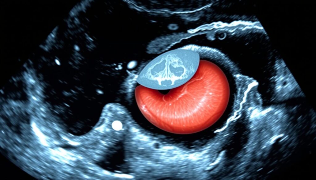
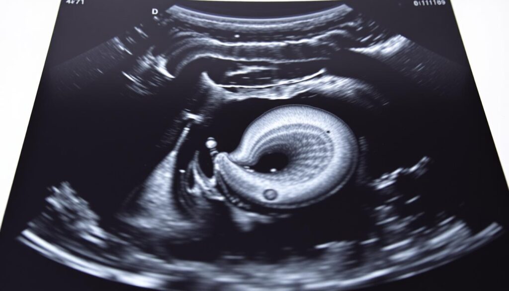
Abdominal aortic aneurysm (AAA) ultrasound is a key tool for finding and tracking aneurysms. At Liv Hospital, we stress the need for precise aorta measurements. This is for effective risk assessment and treatment planning.
An abdominal aortic aneurysm is often found by chance during a physical exam or imaging test. To spot an aneurysm, a doctor checks the aorta’s size. They look at a transverse measurement of 3.0 cm or more as a sign of an aneurysm.
We show you the 7 essential steps for accurate aorta measurement and image interpretation with AAA ultrasound. This ensures top-notch care and exact diagnosis.

Abdominal aortic aneurysms (AAAs) are a serious vascular condition. They need to be found and treated quickly. As healthcare workers, knowing how important AAAs are helps us care for our patients better.
An abdominal aortic aneurysm is when the aorta in the belly gets too big, bigger than 3 cm. More older adults have AAAs, and men over 65 who smoked should get checked by ultrasound. Men who never smoked might need a check too, depending on their family history.
Checking for AAAs by touch alone isn’t enough. Most AAAs don’t show symptoms until they burst. That’s why regular checks are key to finding them early. The US guidelines say screening can cut down deaths from burst aneurysms.
Ultrasound is a key tool in finding AAAs. It’s shown to greatly lower death rates from aneurysms. So, using ultrasound in screenings is very important.
AAA ultrasound is key in vascular imaging. It’s very sensitive and specific. We use it to give accurate diagnoses and plans for patients with abdominal aortic aneurysms.
One big plus of AAA ultrasound is its ability to measure aneurysm size accurately. Ultrasound technology gives precise measurements. These are key for figuring out how serious the aneurysm is and what treatment is needed. Also, Doppler ultrasound checks blood flow in the aorta. This gives important info about how the aneurysm affects blood flow.
AAA ultrasound is very good at detecting aneurysms and tracking their growth. Its high sensitivity and specificity make it a top choice for diagnosis. This accuracy helps us know who needs surgery and who doesn’t.
AAA ultrasound is also budget-friendly for screening and monitoring. It’s cheaper than CT or MRI and doesn’t use radiation. This makes it a great choice for regular checks and long-term monitoring of aneurysms.
Using AAA ultrasound, we can give our patients accurate diagnoses and treatment plans. This technology is essential in our mission to provide top-notch healthcare.
The USPSTF gives advice on when to screen for AAA. They aim to catch problems early and help people get better care.
The USPSTF says men aged 65–75 who smoked should get a one-time ultrasound. This helps lower the risk of death from AAA.
Some people should get screened for AAA sooner. This includes those with a family history of AAA and smokers. They need to be checked earlier than others.
| Risk Factor | Screening Recommendation |
|---|---|
| Men aged 65-75 who have ever smoked | One-time AAA ultrasound screening |
| Family history of AAA | Early screening |
| Smoking history | Early screening |
Following these guidelines helps doctors find and treat AAA early. This can save lives.
To get the best AAA ultrasound images, it’s key to set up the ultrasound right and position the patient well. Having the right equipment and preparation is vital for accurate diagnosis.
Choosing the right ultrasound probe and settings is very important for clear images of the abdominal aorta. A curvilinear probe with a frequency of 3-5 MHz is usually best for abdominal ultrasounds. Adjust the depth and gain to see the aorta and nearby structures clearly.
How the patient is positioned is key to avoiding bowel gas interference. Patients lie on their backs, and the sonographer might use gentle pressure or have them breathe deeply. This helps move bowel gas and improves aorta visibility.
Experts say, “Optimal patient preparation and ultrasound technique are essential for accurate measurement of the aorta on ultrasound”
“The quality of the ultrasound examination is highly dependent on the skill of the sonographer and the quality of the equipment used.”
With the right equipment and patient setup, healthcare providers can get high-quality AAA ultrasound images for accurate diagnosis.
Finding the abdominal aorta is the first step in checking for aneurysms. It’s key for taking precise measurements and understanding the ultrasound images. This step is essential for a thorough AAA ultrasound.
We start by tracing the abdominal aorta from the diaphragm to where it splits into the common iliac arteries. Using ultrasound, we follow it from the diaphragm’s opening. Getting the right angle and position of the transducer is very important for a clear view.
It’s important to tell the aorta apart from other structures. The IVC is usually on the right and has thinner walls than the aorta. Vertebral shadows might look like the aorta but show up as bright lines with shadows behind them. Pay close attention to these landmarks for accurate identification.
| Structure | Location | Ultrasound Characteristics |
|---|---|---|
| Aorta | Midline, slightly left | Pulsatile, thick walls |
| IVC | Right of the aorta | Collapsible, thin walls |
| Vertebral Bodies | Posterior to aorta | Hyperechoic with posterior shadowing |
Learning these techniques helps doctors accurately find the abdominal aorta. This is a critical step in the AAA ultrasound process.
Learning the transducer technique is key for clear aortic views during ultrasound exams. This skill helps us get accurate aorta measurements and make the right diagnoses.
We use both transverse and longitudinal scans to see the aorta well. The transverse approach helps us check the aorta’s size and spot aneurysms. The longitudinal approach shows the aorta’s length and any issues like thrombi or wall problems.
“Using different scanning planes is vital for a detailed aorta check,” say ultrasound experts. Mixing these methods gives us a full picture of the aorta’s shape.
For obese patients, we need to adjust pressure and angle to see the aorta better. We use gentle pressure to move bowel gas and adjust the angle to fit the patient’s body.
By getting better at these techniques, we can make our images clearer and our diagnoses more accurate. A study on ultrasound methods points out, “the right transducer placement and adjustments are essential for top-notch images.”
To get accurate AAA results, it’s key to follow a set measurement plan. Measuring from outer wall to outer wall in both directions is vital. This helps in deciding the best treatment.
The outer wall to outer wall method is a top choice for AAA scans. It measures the aorta’s diameter from the outside of the front wall to the back wall. Being consistent in how you measure is important for getting reliable results.
Things like bowel gas, obesity, and wrong transducer placement can mess up AAA measurements. To avoid these, use a clear imaging plan. This includes the right patient position and adjusting the ultrasound settings.
Experts say paying close attention to details is essential for AAA ultrasound scans. By sticking to a standard protocol and knowing common mistakes, we can get accurate measurements. These measurements help doctors make better choices for patients.
The fourth step in our OCT-5595-AAA Ultrasound protocol is to take diagnostic images in many planes. This helps us understand the aorta fully. It’s key for spotting any issues with the aortic anatomy.
It’s important to take images from both sides and lengths. Transverse images show the aorta’s size and any bulges. Longitudinal images tell us about the aorta’s length and any blockages or hardening.
| Plane | Assessment Focus | Diagnostic Value |
|---|---|---|
| Transverse | Aortic diameter, aneurysms | Detection of irregularities |
| Longitudinal | Aortic length, thrombi, wall calcifications | Assessment of aortic morphology |
For tricky parts of the aorta, we adjust the probe angle and use Doppler ultrasound. This makes the images clearer. It helps us get precise aorta ultrasound images and ultrasound aorta measurements.
By taking images from different angles and using special techniques, we get a full view of the abdominal aorta. This detailed look is essential for making accurate diagnoses and treatment plans.
The fifth step in our OCT-5595-AAA ultrasound protocol is to analyze the aortic morphology and any associated pathology. This step is key to understanding the aneurysm’s complexity and planning the right management.
Intraluminal thrombus and wall calcification are important factors. They can affect the risk of rupture and the management plan. Intraluminal thrombus is blood clots inside the aneurysm sac, seen as hypoechoic or heterogeneous material on ultrasound. Wall calcification is hardened plaque on the aortic wall, seen as bright echoes with acoustic shadowing.
Understanding the aneurysm’s shape, extension, and branch involvement is vital. It helps in planning surgery and assessing risks. The shape, whether fusiform or saccular, affects the risk of rupture.
| Aneurysm Characteristic | Ultrasound Findings | Clinical Implication |
|---|---|---|
| Intral and Thrombus | Hypoechoic or heterogeneous material within the aneurysm sac | Influences rupture risk and management planning |
| Wall Calcification | Bright echoes with acoustic shadowing | Indicates hardened plaque; affects wall integrity |
| Aneurysm Shape | Fusiform or saccular | Impacts rupture risk assessment |
By analyzing these factors, we can fully understand the aneurysm. This helps us develop a suitable treatment plan.
Understanding AAA ultrasound measurements is key. We need to know about aneurysmal criteria and risk stratification. This helps us manage patient care based on aneurysm size and its risks.
A measurement of 3.0 cm or more is seen as an abdominal aortic aneurysm. Accurate measurement is critical for making decisions. It helps us tell normal aortic sizes from aneurysms.
Risk levels depend on aneurysm size, divided into three groups: under 3.0 cm, 3.0-5.4 cm, and ≥5.5 cm. Each group has different implications for patient care. For example, small aneurysms under 3.0 cm might not need immediate action. But large ones, over 5.5 cm, might need surgery.
We categorize risk this way:
Knowing these categories helps us make better decisions for patient care.
Creating detailed reports is key in AAA ultrasound assessments. We make sure all important info is documented well. This info is then shared with healthcare providers.
A good AAA ultrasound report should have all the details. It should talk about the aneurysm’s size and shape. It should also mention any blood clots or wall calcification. Accurate measurement of the aneurysm’s size is very important. It helps doctors decide on the best course of action.
It’s very important to share critical findings quickly. We have set protocols to make sure critical results are shared fast with the doctor or healthcare team.
In conclusion, making detailed reports and documents is a big part of AAA ultrasound assessments. By including all the important details and following our communication rules, we help patients get the care they need.
Accurate aorta measurement and image interpretation are key for managing abdominal aortic aneurysms (AAA). By following the 7 key steps in this article, healthcare professionals can excel in AAA assessment and patient care.
We aim to offer top-notch healthcare to international patients. This includes precise AAA ultrasound assessments. This helps in timely interventions and better patient outcomes.
Excellence in AAA care means more than just accurate diagnosis. It also includes risk stratification, monitoring, and referrals for surgery or endovascular interventions. This approach optimizes patient care and lowers the risk of rupture.
Effective AAA assessment and patient management need a team effort. Radiologists, vascular surgeons, and other experts work together. They use the latest ultrasound technologies to improve patient care and outcomes.
Our dedication to excellence in AAA assessment and patient management can change lives worldwide.
The normal diameter of the abdominal aorta is usually under 2.0 cm. A diameter of 3.0 cm or more is considered an aneurysm.
Ultrasound is key in finding abdominal aortic aneurysms. It’s very sensitive and specific. Plus, it’s affordable for screenings and follow-ups.
The USPSTF suggests screening men aged 65-75. People with risk factors should also get screened early.
To find the abdominal aorta, landmarks are used. It goes from the diaphragm to where it splits. It’s also distinguished from the IVC and vertebral shadows.
To master transducer placement, use both transverse and longitudinal scans. Adjust pressure and angle, which is important for obese patients.
The size is measured from the outer wall to outer wall. It’s important to avoid common mistakes in measurement.
Images in both transverse and longitudinal planes are key. They help document and visualize the aorta fully.
When assessing the aorta, look for intraluminal thrombus and wall calcification. Also, check the aneurysm’s shape, extension, and branch involvement.
Measurements are interpreted based on size. Sizes under 3.0 cm, 3.0-5.4 cm, and over 5.5 cm are risk-stratified.
An AAA ultrasound report should detail findings clearly. It should also have protocols for urgent findings.
Measuring aorta size is vital for detecting and monitoring AAA. It helps in making timely decisions.
Ultrasound aids in screening, surveillance, and monitoring AAA. It provides essential information for managing the condition.
Subscribe to our e-newsletter to stay informed about the latest innovations in the world of health and exclusive offers!
WhatsApp us