Last Updated on November 27, 2025 by Bilal Hasdemir

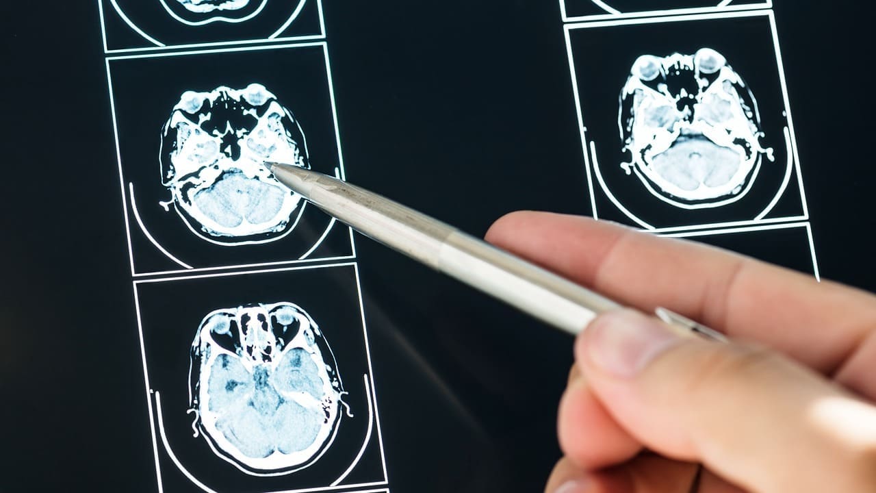
Diagnosing traumatic brain injuries and concussions right is key for good care later on. At Liv Hospital, we know how vital it is to spot past damage. That’s why we use top-notch CT and MRI technology for solid checks.
But, regular scans might miss how bad past damage is. This makes it hard to give the right care. We stick to the latest medical studies and rules to give a full picture to our readers.
We’ll look at what CT and MRI scans can do to find traumatic brain injuries and concussions. We aim to mix medical know-how with a caring touch.
Traumatic brain injuries, including concussions, are a range of conditions. They can affect people differently. These injuries happen when the brain moves inside the skull due to outside forces.
TBI can be mild or severe. The Glasgow Coma Scale (GCS) helps measure how severe it is. Mild TBI, or concussion, usually has temporary symptoms. But severe TBI can cause long-term problems like memory loss and disability.
| TBI Severity | GCS Score | Typical Symptoms |
|---|---|---|
| Mild | 14-15 | Confusion, headache, dizziness |
| Moderate | 9-13 | More pronounced confusion, possible loss of consciousness |
| Severe | 3-8 | Prolonged unconsciousness, significant cognitive impairment |
Right after a TBI, you might feel confused, have a headache, or feel dizzy. But, long-term effects can be different. They might include ongoing brain problems, mood changes, and a higher risk of diseases like Alzheimer’s. Knowing these effects helps doctors and patients better manage TBI.
Understanding neuroimaging is key to assessing brain trauma well. It’s a vital tool in diagnosing and managing traumatic brain injuries (TBI). It helps doctors make the right treatment choices.
Brain imaging has changed a lot over time. We’ve moved from simple X-rays to advanced tools like CT scans and MRI. CT scans are often the first choice for acute TBI. They’re fast and show important details like hemorrhages and fractures.
MRI gives detailed views of the brain. It’s great for spotting small injuries and seeing how much damage there is, even in later stages of TBI.
Imaging is needed after head trauma if there’s a big injury. This includes losing consciousness, having a severe headache, or a penetrating head injury. The choice between CT and MRI depends on the situation.
CT scans are best in emergency situations because they’re quick and show bleeding well. MRI is used later to see the full injury. It helps in planning rehabilitation.
We use neuroimaging to diagnose and track brain injuries. It helps us understand how to help patients with TBI better.
It’s important to know what CT scans can and can’t do when it comes to brain injuries. CT scans are often used in emergencies to check for head trauma. But, they’re not always good at finding injuries that happened a long time ago.
CT scans are great at finding injuries right after they happen. They can spot bleeding, broken bones, and other immediate problems. They work well because they can see bone and fresh blood clearly. This is key in emergencies when doctors need to act fast.
But, CT scans aren’t as good at finding injuries that happened a while ago. Over time, injuries can look different on a CT scan, making them harder to see. Small injuries or damage to brain tissue might not show up. This means some people with old brain injuries might look fine on a CT scan, even if they’re not feeling well.
Even though CT scans have their limits, they’re very useful in emergency situations. Knowing what they can and can’t do helps doctors make better choices for their patients. This includes when to use other tests like MRI to get a full picture of what’s going on.
Getting a normal CT scan result after a head injury can be a big relief. But, it’s important to know what this really means. A normal result doesn’t always mean there’s no brain injury.
Studies show that up to 80% of traumatic brain injuries (TBIs) might not show up on CT or MRI scans. This doesn’t mean the injuries are not serious. It shows how limited current imaging is in finding some brain damage.
Invisible injuries include tiny damage to brain cells or changes in brain function.
People with normal CT scans after head injuries might have symptoms like headaches or trouble thinking. This shows we need more than just scans to check for injuries. We also need clinical checks and advanced imaging.
A normal CT scan is just one part of figuring out head injuries. We might need more tests to really understand the damage.
MRI technology has changed how we detect brain injuries. It gives us detailed views of the brain’s damage. This helps doctors know how bad the injury is and plan the best treatment.
Standard MRI scans are often the first choice for brain injury imaging. They show the brain’s structures clearly. But, advanced MRI techniques like DTI and fMRI reveal more about the brain’s inner workings. They spot changes that standard scans might miss.
Research shows MRI can find signs of old brain injuries, even if symptoms are gone. Studies have shown advanced MRI can spot changes and markers of past brain injuries.
We use the newest MRI tech to make accurate diagnoses and plan treatments for brain injury patients. By mixing standard and advanced MRI, we get a full picture of each patient’s condition.
It’s important to know the difference between concussion and TBI for the right treatment. Both are serious injuries that affect people in different ways.
A concussion is a mild form of TBI. It has symptoms that usually go away on their own. But, getting hit in the head too many times can cause lasting problems.
Imaging is key in figuring out how bad a brain injury is. New imaging methods can spot small changes in the brain. These changes help tell if a TBI is mild or more serious.
| Imaging Marker | Mild TBI (Concussion) | Moderate/Severe TBI |
|---|---|---|
| CT Scan Findings | Often Normal | May show hemorrhage, edema |
| MRI Findings | May show microstructural changes | Often shows significant structural damage |
Imaging findings are key in figuring out how bad a TBI is. They help doctors decide the best treatment. We use different imaging methods to see the full extent of the injury.
When we use MRI to check for traumatic brain injuries (TBI), we look for specific signs. MRI gives us detailed images of the brain. This helps us find any damage.
One important thing we look for is microhemorrhages. These are small bleeds in the brain. They show how severe the TBI is. We also check the white matter tracts for damage. Microhemorrhages and white matter changes are critical markers of TBI.
For older injuries, MRI shows scarring and atrophy. Scarring looks like gliosis or encephalomalacia. Atrophy means less brain tissue. These signs tell us about the long-term effects of TBI. We study them closely to understand the injury’s extent.
It’s key to match the MRI findings with the patient’s symptoms. We don’t just look at the images. We also consider the patient’s overall health. This helps us understand how the injury affects the brain and overall health.
By combining MRI scans with clinical data, we can make a more accurate diagnosis. This helps us create a good treatment plan. This approach is vital for managing TBI patients.
For patients with suspected traumatic brain injuries, CT scans are a fast and reliable way to find injuries that need quick medical care. We use CT scans first because they can quickly spot serious problems like bleeding, fractures, and other dangers.
CT scans are often the first choice for acute traumatic brain injuries. They are fast and available, even in emergencies. This quickness is key for diagnosing and treating TBI patients right away.
After the first scan, we might suggest more imaging with CT or MRI. This depends on how the patient is doing and what the first scan showed. If there’s an injury, more scans might be needed to watch how it changes or to find new injuries.
During a CT scan, patients lie on a table that moves into the scanner. The whole thing takes just a few minutes. We make sure patients are comfortable and know what’s happening to help them feel less stressed.
By sticking to the best CT scan practices for TBI, we help patients get the right diagnosis fast. This leads to better treatment and care for them.
The way we diagnose traumatic brain injuries (TBIs) is changing. New technologies are helping us see old brain injuries better than before.
Diffusion Tensor Imaging (DTI) is a high-tech MRI method. It shows how well white matter tracts in the brain work. DTI is great for spotting small changes after a TBI, even if regular MRI scans look fine.
It looks at how water moves in nerve fibers. This helps find damage or changes that show old injuries.
Functional MRI (fMRI) and Positron Emission Tomography (PET) scanning are advanced tools. They help us understand brain function and how it uses energy. fMRI tracks blood flow to see brain activity. PET uses special tracers to show how cells work.
Both help find where old injuries are in the brain. This info is key for figuring out what treatments to use.
New tools are coming to help find TBIs better. Things like magnetic resonance spectroscopy (MRS) and susceptibility-weighted imaging (SWI) might give us more clues. As these tools get better, we’ll learn more about TBIs and their lasting effects.
| Neuroimaging Technique | Primary Use in TBI Detection | Key Benefits |
|---|---|---|
| DTI | Assessing white matter integrity | Detects microstructural changes, reveals axonal injury |
| fMRI | Mapping brain activity | Identifies functional changes, aids in treatment planning |
| PET Scanning | Visualizing metabolic processes | Provides insights into brain metabolism, detects areas of injury |
Testing for traumatic brain injuries (TBI) years later is complex. It goes beyond just looking at images. We use a mix of clinical checks, patient history, and new diagnostic tools to see how TBI affects people long-term.
Testing for TBI involves many steps. We check how well the brain works, memory, and emotions. We also look at the patient’s health history, symptoms, and overall well-being.
Imaging like MRI and CT scans is helpful. But, matching these images with the patient’s symptoms is key. This helps us see how much the injury has affected them.
Case studies show how tricky it can be to diagnose old TBI. For example, someone who had a concussion years ago might keep having problems. New tests and careful checks can find the cause.
| Case | Symptoms | Imaging Findings | Diagnosis |
|---|---|---|---|
| 1 | Memory loss, headaches | Microhemorrhages on MRI | Old TBI |
| 2 | Dizziness, cognitive fog | White matter changes on DTI | Traumatic brain injury sequelae |
Liv Hospital leads in diagnosing brain injuries with the latest imaging methods. We stick to international standards to ensure top-notch care. Our dedication to excellence shows in our strict adherence to global neuroimaging best practices.
We adhere to global brain injury imaging protocols. This means our patients get diagnoses backed by the latest research and guidelines. It helps us give accurate and trustworthy brain injury assessments.
We use advanced imaging methods like Diffusion Tensor Imaging (DTI) and Functional MRI. These tools help us see brain injuries more clearly. They give us a deeper look at how injuries affect the brain.
| Imaging Technique | Application in Brain Injury |
|---|---|
| Diffusion Tensor Imaging (DTI) | Assesses white matter tracts and detects microstructural changes |
| Functional MRI | Evaluates brain activity and identifies areas affected by injury |
Our advanced imaging results help shape treatment plans. This way, our doctors can create targeted and effective care. By linking imaging findings with symptoms, we offer complete care for our patients.
Detecting old brain injuries and concussions is a complex task. Advanced neuroimaging techniques are needed. CT and MRI scans are key in showing the extent of brain damage. MRI is very good at spotting old brain injuries.
The future looks bright for detecting old brain injuries. New imaging technology is on the horizon. Improvements in MRI, like diffusion tensor imaging and functional MRI, will help us better detect and manage these injuries. This will lead to better care and outcomes for patients.
At Liv Hospital, we’re all about top-notch healthcare with the latest tech. Our team is ready to give international patients, including those with brain injuries, the best care. With new imaging tech, we’ll be able to help our patients even more, improving their lives.
A concussion is a mild form of TBI. People often use these terms together. But TBI covers a wide range of brain injuries, from mild to severe. The severity of the injury affects what imaging can show.
CT scans are not great at finding older brain injuries. They work best in the first moments after a trauma. They can spot hemorrhages and fractures quickly. But, they might miss older injuries.
A normal CT scan after a head injury doesn’t always mean there’s no injury. Some injuries are too small for a CT scan to see. This shows we need more than just imaging to check for injuries.
Yes, MRI can find old brain injuries. Advanced MRI can spot tiny changes in the brain. These changes might show up years after an injury, like microhemorrhages or scarring.
Testing for TBI years later needs a full approach. This includes checking the patient’s history and doing imaging studies. It’s important to match what the scans show with the patient’s symptoms.
Advanced imaging like Diffusion Tensor Imaging (DTI) and PET scanning help find old brain injuries. These tools give detailed views of the brain. They help doctors decide on the best treatment.
CT scans are often the first choice after head trauma. They’re fast and easy to get. They’re best in emergency situations where quick results are needed.
Radiologists look for signs of injury on MRI scans. They check for microhemorrhages and scarring. They also match what they see with the patient’s symptoms to understand the injury fully.
Imaging results are key in deciding how to treat TBI. They show how severe the injury is. This helps doctors create a treatment plan that fits the patient’s needs.
Subscribe to our e-newsletter to stay informed about the latest innovations in the world of health and exclusive offers!