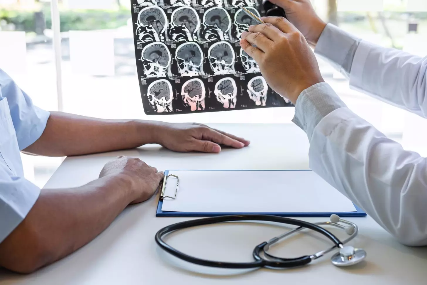
Magnetic resonance imaging (MRI) is key for spotting brain tumors, like glioblastoma. At Liv Hospital, we use top-notch MRI tech for precise diagnoses and treatment plans for our patients.
MRI scans help doctors see what brain cancer looks like and its key features. We use this info to plan treatments and give top-notch care to brain cancer patients.
We mix trusted expertise with a patient-centered, team approach. This way, we understand MRI images to find out what the mass is and create a treatment plan just for you.
Key Takeaways
- MRI is a vital diagnostic tool for detecting brain tumors.
- Glioblastoma has distinct characteristics on MRI scans.
- Liv Hospital uses advanced MRI technology for accurate diagnoses.
- A multidisciplinary approach ensures effective treatment plans.
- Personalized care is provided to each patient based on their MRI results.
Understanding MRI Technology for Brain Imaging

Advanced MRI technology is key in seeing brain tissue and finding problems. MRI, or Magnetic Resonance Imaging, uses strong magnetic fields and radio waves. It creates detailed images of the brain’s inside.
How MRI Works to Visualize Brain Tissue
MRI machines align hydrogen atoms in the body with a strong magnetic field. Radio waves disturb these atoms, causing them to send signals. The MRI machine catches these signals and makes images.
This process is safe because it doesn’t use radiation. It’s also non-invasive, making it safe for patients.
The clarity of MRI images lets doctors see different brain tissues. This is key for finding brain cancer and planning treatment.
“MRI has revolutionized the field of neuroimaging, providing unparalleled detail of brain structures and pathology.” –
Types of MRI Sequences Used in Brain Cancer Detection
There are different MRI sequences for different brain tissue features. T1-weighted images show anatomy well. T2-weighted images are better for finding tumors.
- T1-weighted images with contrast enhancement
- T2-weighted images for detecting edema and lesions
- FLAIR sequences for suppressing fluid signals
- Diffusion-weighted imaging for assessing tumor cellularity
Doctors use these sequences to get full info on brain tumors. They can see size, location, and more.
The Basics of Brain Cancer Visualization on MRI

Knowing how brain cancer looks on MRI is key for accurate diagnosis and treatment planning. MRI gives us detailed images of brain tumors. This helps us see their characteristics and plan the best treatment.
General Characteristics of Brain Tumors on MRI
Brain tumors show up differently on MRI, depending on their type and grade. Usually, they look like a mass with an irregular shape and mixed signals. It’s important to know these general traits.
- Irregular shape and poorly defined borders
- Heterogeneous signal intensity
- Variable enhancement patterns after contrast administration
- Mass effect and surrounding edema
Differentiating Cancerous vs. Non-Cancerous Lesions
Telling cancerous from non-cancerous lesions is a big part of diagnosis. We use MRI sequences to make this distinction. Cancerous lesions often grow fast and spread into nearby tissues.
| Feature | Cancerous Lesions | Non-Cancerous Lesions |
|---|---|---|
| Growth Pattern | Rapid, invasive | Slow, expansive |
| Border Definition | Poorly defined | Well-defined |
| Contrast Enhancement | Heterogeneous, often strong | Homogeneous, variable |
Common Signs of Malignancy in Brain Imaging
When we check brain tumors on MRI, we look for signs of cancer. These include irregular shape, mixed signals, and big mass effect. Also, seeing necrosis in a tumor is a red flag.
By knowing these signs, we can better diagnose and treat brain cancer. This helps improve patient results.
What Does Brain Cancer Look Like on MRI: Key Imaging Features
MRI scans show specific signs of brain cancer that are key for correct diagnosis. When looking at brain cancer on MRI, several important features can be seen. These help tell it apart from other conditions.
Irregular Shape and Poorly Defined Borders
Brain cancer on MRI looks like a mass with an irregular shape and unclear borders. This irregular shape is a sign of cancer because it spreads into the brain tissue around it.
Glioblastoma, a very aggressive brain cancer, often has an irregular shape on MRI. Its borders are hard to see, making it hard to know how big the tumor is.
Heterogeneous Enhancement Patterns
Another important sign of brain cancer on MRI is the heterogeneous enhancement pattern. After getting contrast agents like gadolinium, cancer tumors show different enhancement patterns.
For example, glioblastoma often has a mixed enhancement pattern with dead areas and uneven enhancement. This is because the tumor has different parts like fast-growing areas, dead spots, and blood vessel growth.
“The heterogeneous enhancement pattern observed in glioblastoma is a result of its aggressive nature and complex vascular structure.”
Mass Effect and Surrounding Edema
Brain cancer can also cause a mass effect, pushing brain structures away. This can lead to different symptoms depending on where the tumor is.
Also, edema, or swelling, is often seen around brain cancer. Edema is fluid buildup in the brain tissue near the tumor. On MRI, it looks bright on T2-weighted images.
| Imaging Feature | Description | Clinical Significance |
|---|---|---|
| Irregular Shape | Tumor with irregular borders | Indicative of malignancy |
| Heterogeneous Enhancement | Varied enhancement patterns after contrast | Suggests tumor complexity and aggressiveness |
| Mass Effect | Displacement of surrounding brain structures | Can cause neurological symptoms |
| Surrounding Edema | Swelling around the tumor | Contributes to mass effect and neurological symptoms |
Knowing these key features of brain cancer on MRI is vital for accurate diagnosis and treatment planning. By recognizing these signs, doctors can better diagnose and manage brain cancer.
Glioblastoma MRI Images: The Most Aggressive Brain Cancer
Glioblastoma is the most aggressive brain cancer. It shows unique signs on MRI images that help doctors diagnose it. MRI helps us see how big the tumor is and plan treatment.
Characteristic Appearance of Glioblastoma
Glioblastoma looks like a lesion with thick, irregular edges and a dead center on MRI. This is because the tumor grows fast and outgrows its blood supply, causing death of cells.
Ring-Like Enhancement and Central Necrosis
Glioblastoma is known for its ring-like enhancement around a dead center on MRI. This is seen after using a contrast agent like gadolinium. It shows the tumor’s blood flow and structure. A study on the National Center for Biotechnology Information website says this pattern is key for diagnosing glioblastoma.
Stage 4 Glioblastoma MRI Findings
At Stage 4, glioblastoma shows aggressive signs on MRI. These include a big mass, swelling around it, and uneven enhancement. The tumor can also spread to nearby brain areas, making surgery hard.
| MRI Feature | Description | Clinical Significance |
|---|---|---|
| Ring-like Enhancement | Enhancement around a necrotic center | Indicative of glioblastoma’s aggressive nature |
| Central Necrosis | Dead tissue within the tumor | Result of rapid tumor growth outpacing blood supply |
| Heterogeneous Enhancement | Varied appearance on MRI due to different tumor components | Reflects tumor’s complex structure and aggressiveness |
Tumor in Brain MRI Images: Location Matters
The spot where a tumor grows in the brain changes how it looks on MRI scans. Knowing where a tumor is helps doctors diagnose and plan treatment. We’ll look at how a tumor’s location affects its MRI look and where different brain cancers usually grow.
Supratentorial vs. Infratentorial Tumors
Brain tumors are split into two groups based on where they are in the brain. Supratentorial tumors are above a certain membrane, in the cerebrum. Infratentorial tumors are below it, in the cerebellum or brainstem. This matters because it can tell doctors what kind of tumor it is and how it might act.
Adults usually get supratentorial tumors, which can come from different brain cells. Kids often get infratentorial tumors, which are more likely to be in the cerebellum or fourth ventricle.
How Location Affects MRI Appearance
The look of brain tumors on MRI scans changes based on their location. Tumors in different spots can look different because of the brain’s structure and what it’s made of. For example, tumors near the ventricles might look different than those in the outer brain.
The brain around a tumor can swell, which MRI can show. How much swelling there is and what it looks like can tell doctors about the tumor’s growth and type.
Common Locations for Different Types of Brain Cancer
Brain cancers often grow in certain areas. For example, glioblastomas usually grow in the brain’s hemispheres, like the frontal or temporal lobes. Meningiomas grow from the brain’s protective membranes and are often near the brain’s surface.
Knowing where tumors usually grow helps doctors figure out what kind they are. This lets them make better diagnoses and treatment plans by matching the tumor’s location with its MRI look.
Contrast Enhancement in Brain Cancer MRI
Gadolinium-based contrast agents have greatly improved MRI images of brain tumors. They help see tumors clearly, making diagnosis and treatment planning more accurate.
The Role of Gadolinium in Brain Tumor Imaging
Gadolinium is a contrast agent that makes brain tumors visible on MRI. It builds up in areas where the blood-brain barrier is broken, like around tumors. This makes the tumor stand out on the MRI scan.
We use gadolinium-based contrast agents to:
- Enhance the visibility of tumor boundaries
- Differentiate between tumor types
- Assess tumor vascularity
Patterns of Enhancement in Different Types of Brain Cancer
Brain cancers show different enhancement patterns on MRI. For example:
| Tumor Type | Typical Enhancement Pattern |
|---|---|
| Glioblastoma | Ring-like enhancement with central necrosis |
| Meningioma | Homogeneous enhancement |
| Metastatic Tumors | Variable, often ring-like or nodular enhancement |
Non-Enhancing Tumors and Their Significance
Not all brain tumors show up with gadolinium. Non-enhancing tumors are just as important. They might have a closed blood-brain barrier or be less vascular. Knowing about these tumors is key for correct diagnosis and treatment.
When looking at MRI scans for brain cancer, we must consider both types. This detailed approach helps get a precise diagnosis and the best treatment plan.
Peritumoral Changes and Edema on MRI
Understanding peritumoral changes and edema is key for accurate brain tumor assessment on MRI. Peritumoral changes are the changes in brain tissue around a tumor. These changes can affect diagnosis and treatment planning. Edema is a common change, where too much fluid builds up in brain tissue.
Vasogenic Edema Surrounding Brain Tumors
Vasogenic edema happens when the blood-brain barrier breaks down, often due to tumors. It shows up as bright spots on T2-weighted MRI sequences around the tumor. The amount of vasogenic edema depends on the tumor’s type and how aggressive it is.
Research shows vasogenic edema is more common in aggressive tumors like glioblastoma than in less aggressive ones (PMC4358863). A lot of vasogenic edema can mean the tumor is growing fast.
Differentiating Tumor Infiltration from Edema
Telling tumor infiltration apart from edema is important for accurate diagnosis and treatment. Tumor infiltration means cancer cells spreading into brain tissue. Edema is just too much fluid.
- Tumor infiltration looks more uniform on MRI.
- Edema looks more mixed up.
- Advanced MRI methods like diffusion-weighted imaging and MR spectroscopy can tell them apart.
The Significance of Peritumoral Signal Abnormalities
Peritumoral signal abnormalities on MRI offer clues about the tumor’s aggressiveness and how it might spread. These can include changes in signal, mass effect, and edema.
Key features to consider:
- The extent of peritumoral edema and its impact on surrounding brain structures.
- The presence of tumor infiltration into adjacent brain tissue.
- The overall mass effect caused by the tumor and associated edema.
By closely looking at peritumoral changes and edema on MRI, doctors can better understand the tumor. This helps them plan more effective treatments.
Advanced MRI Techniques for Brain Cancer Visualization
Advanced MRI techniques have changed how we see brain cancer. They give us detailed views of tumors. This helps us diagnose and treat brain cancers better.
We use many advanced MRI techniques to understand brain cancer. Some key methods include:
Diffusion-Weighted Imaging (DWI)
Diffusion-Weighted Imaging (DWI) measures water molecule movement in tissues. It shows where water can’t move much, which might mean tumor cells or dead tissue.
Key benefits of DWI in brain cancer visualization:
- Helps tell different tumor types apart by looking at cell density
- Finds areas of dead tissue or lack of blood flow in tumors
- Gives important info for planning and checking on treatments
Perfusion MRI
Perfusion MRI looks at blood flow and how tissues take it in. It’s great for seeing how blood-rich tumors are and how they change over time.
The info from Perfusion MRI is very useful in:
- Seeing how aggressive a tumor is by looking at blood flow
- Watching how well treatments that stop blood flow to tumors work
- Telling the difference between a new tumor and damage from treatment
MR Spectroscopy
MR Spectroscopy is a way to look at brain tissue’s chemistry without surgery. It can tell us what kind of brain lesions we’re dealing with and how treatments are working.
MR Spectroscopy can help find:
- Chemical signs of how aggressive a tumor is
- Changes in tumor chemistry after treatment
- How to tell if a new growth is a tumor or treatment damage
By using these advanced MRI techniques together, we can understand brain cancer better. This leads to more accurate diagnoses and better treatment plans.
The Diagnostic Process: From MRI Images to Brain Cancer Diagnosis
Diagnosing brain cancer is a detailed process. It involves looking at MRI images, symptoms, and biopsy results. We will explain the steps from the start to the diagnosis.
Radiological Assessment and Reporting
Looking at MRI images is key in diagnosing brain cancer. We check the tumor’s size, location, and how it looks on the scan. Our advanced imaging helps us give a detailed report for treatment planning.
The report tells about the tumor’s shape, how it affects nearby areas, and if it’s near important brain parts. This info helps doctors decide the best treatment.
Correlation with Clinical Symptoms
Matching MRI images with symptoms is vital. We look at symptoms like headaches and seizures to see how the tumor affects the brain. This helps us understand how aggressive the tumor is.
We also check the patient’s medical history and other tests to rule out other causes. By combining MRI findings with symptoms, we can make a more accurate diagnosis.
“The correlation between imaging findings and clinical presentation is key in diagnosing brain tumors. It needs a team effort from neuroradiologists, neurosurgeons, and oncologists for the best care.”
The Role of Biopsy in Confirming MRI Findings
Even with MRI, a biopsy is often needed to confirm the diagnosis. We take tissue samples for histopathological examination. This tells us the tumor type and grade.
| Diagnostic Step | Purpose | Outcome |
|---|---|---|
| MRI Imaging | Visualize tumor characteristics | Identify tumor location, size, and enhancement patterns |
| Clinical Correlation | Assess tumor impact on brain function | Understand symptom severity and tumor aggressiveness |
| Biopsy | Confirm tumor type and grade | Histopathological diagnosis |
The process of diagnosing brain cancer is complex. By combining MRI images, symptoms, and biopsy results, we can make an accurate diagnosis. This helps us create a treatment plan that meets the patient’s needs.
Differentiating Brain Cancer from Other Conditions on MRI
It’s key to tell brain cancer apart from other brain disorders on MRI for good treatment plans. We need to closely look at MRI images to spot tumors from other look-alikes.
Brain Abscess vs. Tumor
Brain abscesses and tumors can look alike on MRI. But, abscesses have a clear wall and show certain signs on DWI sequences. Tumors have messy edges and grow differently. Symptoms like fever help tell them apart.
Stroke vs. Tumor
Strokes and tumors can look similar on MRI, but strokes happen fast. They show clear signs on DWI and ADC maps. Tumors grow slower and don’t show the same signs.
Multiple Sclerosis vs. Tumor
MS lesions can look like tumors, but they’re usually near the ventricles. They might show up on contrast scans but not as much as tumors. Tumors take up more space and look different.
Radiation Necrosis vs. Tumor Recurrence
Radiation necrosis can look like new tumors after treatment. It has swelling around it. MRI tests like spectroscopy and perfusion imaging can tell them apart by looking at how they work.
In summary, telling brain cancer from other conditions on MRI needs skill, careful looking, and sometimes more tests. Knowing how different brain issues look on MRI helps us treat patients better.
Conclusion: The Critical Role of MRI in Brain Cancer Detection and Treatment
MRI is key in finding and treating brain cancer. At Liv Hospital, we use MRI to spot brain tumors and plan treatments. MRI’s detailed images help our team accurately diagnose and tailor treatments.
MRI has changed how we fight brain cancer. It shows brain tissue clearly, helping doctors tell cancerous from non-cancerous areas. This is vital for choosing the right treatment for brain cancer patients.
We aim to offer top-notch healthcare, supporting patients from around the world. Our advanced MRI and skilled team ensure patients get the best care for diagnosing and treating brain cancer.
What does brain cancer look like on an MRI?
On an MRI, brain cancer shows up as a mass. It has an irregular shape and uneven enhancement. The look can change based on the tumor’s type and grade.
How is glioblastoma visible on MRI images?
Glioblastoma shows up on MRI with a ring-like enhancement and central necrosis. It looks like a mixed mass with irregular edges and a lot of edema around it.
What is the role of contrast enhancement in brain cancer MRI?
Contrast enhancement, like gadolinium, makes tumors stand out on MRI. It makes them appear brighter. Different cancers show different enhancement patterns.
How do doctors differentiate between brain cancer and other conditions on MRI?
Doctors use MRI sequences, symptoms, and sometimes more tests to tell brain cancer apart from other issues. This includes brain abscess, stroke, multiple sclerosis, or radiation necrosis.
What advanced MRI techniques are used for brain cancer visualization?
Advanced MRI uses techniques like diffusion-weighted imaging (DWI), perfusion MRI, and MR spectroscopy. These give more details about the tumor’s nature and behavior.
How does the location of a brain tumor affect its appearance on MRI?
Where a brain tumor is located can change how it looks on MRI. This is because of the different brain tissue around it. Tumors in different spots can look different.
What is the significance of peritumoral changes and edema on MRI?
Changes and edema around tumors on MRI show how the tumor affects the brain. Knowing this is key for making the right diagnosis and treatment plan.
How is MRI used in the diagnostic process for brain cancer?
MRI is a key tool in diagnosing brain cancer. It gives detailed images that help doctors understand the tumor, plan treatment, and check how well it’s responding to therapy.
Can MRI alone diagnose brain cancer?
MRI is very helpful in diagnosing brain cancer, but it’s not always enough. Doctors often use it with clinical assessment and sometimes biopsy to confirm the diagnosis.
What are the common MRI sequences used for brain cancer detection?
MRI uses sequences like T1-weighted, T2-weighted, FLAIR, and diffusion-weighted imaging. These help show different parts of the tumor and the tissue around it.
How does the appearance of brain cancer on MRI vary between different types of tumors?
Different brain tumors look different on MRI. This depends on the tumor’s grade, location, and what it’s made of.
Reference
- UPenn GBM Collection. The Cancer Imaging Archive. https://www.cancerimagingarchive.net/collection/upenn-gbm
- Glioblastoma, IDH-Wildtype. Radiopaedia. https://radiopaedia.org/articles/glioblastoma-idh-wildtype
- PMC7868590. National Center for Biotechnology Information. https://pmc.ncbi.nlm.nih.gov/articles/PMC7868590


