Last Updated on November 27, 2025 by Bilal Hasdemir

At Liv Hospital, we use brain x-ray imaging to check for skull injuries and brain changes. This method uses ionizing radiation to create images. These images show how different tissues absorb X-rays.
We know how vital x ray brain imaging is for medical checks. Our team of experts is committed to top-notch healthcare for all patients. By learning about X-ray imaging, we can better understand and treat brain issues.
Brain X-ray imaging is key for diagnosing and managing neurological conditions. We’ll look at its role in diagnosing, the types of procedures, and its history.
Brain X-ray imaging is vital for diagnosing neurological disorders. It spots structural issues like fractures or calcifications. These findings help doctors choose the right treatment.
Modern X-ray methods, like CT scans, are essential in emergency and neurological diagnosis.
There are many brain X-ray procedures, each for different uses. Conventional X-rays check the skull’s health. CT scans give detailed brain images.
These tools help find various conditions, from injuries to vascular problems.
The history of brain imaging started with Wilhelm Conrad Röntgen’s X-ray discovery in 1895. Later, CT scans were invented in the 1970s, changing brain imaging forever.
More recent technologies, like PET and MRI, have deepened our understanding of brain issues and blood flow.
Today, CT scans accurately detect brain disorders 78% of the time. They’re used to spot strokes and trauma in nearly half of cases. These advancements keep improving how we diagnose and treat patients.
Skull injuries and abnormalities can be diagnosed with brain X-ray imaging. This tool is key in medical settings, needed in emergencies for quick checks.
We use X-ray technology to look at the brain and skull for different conditions. X-ray imaging’s precision helps spot fractures and other issues.
Brain X-ray imaging is mainly used to find skull fractures. X-ray tech gives a clear view of the skull. This helps doctors spot fractures and judge their severity.
Brain X-ray imaging also shows signs of increased intracranial pressure. This is a serious condition that needs quick diagnosis and treatment.
| Condition | X-Ray Findings | Clinical Significance |
|---|---|---|
| Skull Fracture | Visible fracture line | Indicates trauma, possible intracranial injury |
| Increased Intracranial Pressure | Suture diastasis, pineal gland shift | Life-threatening, needs immediate action |
| Structural Abnormalities | Abnormal calcifications, bony erosions | May show underlying issues, like tumors or infections |
X-ray imaging of the brain shows various skull changes. These signs can point to conditions needing medical care.
Understanding brain X-ray imaging helps doctors make better decisions for patient care.
Traditional brain X-ray imaging is a key tool in diagnosing neurological issues. Yet, it has big limitations. The main issue is its inability to show detailed images of soft tissues.
X-ray technology works by showing differences in tissue density. But soft tissues like the brain are hard to tell apart because they have similar densities. This makes X-ray images often unclear for detailed diagnoses.
Seeing brain tissue with traditional X-rays is tough because of the skull’s dense bones. These bones block the view of soft tissue details. This can lead to unclear diagnoses, needing more tests.
“The introduction of newer imaging techniques like perfusion CT has significantly enhanced our ability to evaluate cerebral vascular physiology and hemodynamics.”
Because of X-ray’s limits, we often need MRI or CT scans. These methods give clearer images of brain tissue and blood vessels. They help in making accurate diagnoses and treatment plans.
| Imaging Modality | Soft Tissue Detail | Diagnostic Use |
|---|---|---|
| Traditional X-Ray | Limited | Skull fractures, foreign objects |
| CT Scan | Moderate | Trauma, hemorrhage |
| MRI | High | Soft tissue abnormalities, tumors |
We know traditional X-rays have their limits but are useful in some cases. Knowing their limits helps us choose better imaging methods when needed.
Modern brain X-ray technologies have changed how we diagnose the brain. They offer deep insights into the brain’s workings. CT scanning has been a big leap forward, changing how we find and treat brain problems.
CT scans are key in brain imaging because they show detailed brain pictures. They help doctors find and fix problems fast. CT angiography has also gotten better, helping doctors more and more.
Research shows CT scans are about 78% accurate in finding brain issues. This high accuracy is vital in emergencies. It helps doctors quickly spot problems like stroke and trauma, improving care.
CT scans are great at spotting stroke, trauma, and other urgent brain issues. They catch these problems fast, making them a top choice in emergencies. For example, they can spot bleeding in the brain quickly, helping doctors act fast.
We count on these advanced brain X-ray tools to give our patients the best care. Knowing what these tools can do helps us make better choices for our patients.
When you get a brain X-ray, you might worry about radiation. This is a big concern for those looking for top-notch medical care.
A brain X-ray exposes you to about 1.05 millisieverts of radiation. The American College of Radiology sets rules to keep radiation use safe. These rules help us get the right images without too much exposure.
We stick to these rules to give our patients the least amount of radiation needed. This way, we can compare different radiation levels.
It’s good to know how much radiation a brain X-ray gives you compared to daily life. For example, you get about 2.4 millisieverts of background radiation each year from nature.
| Radiation Source | Effective Dose (Millisieverts) |
|---|---|
| Brain X-Ray | 1.05 |
| Annual Background Radiation | 2.4 |
| Flight from New York to Los Angeles | 0.1 |
We have strict rules to cut down on radiation. We use the least dose needed and make sure our X-ray machines are in top shape.
We also follow the ALARA principle. This means we always try to use the least amount of radiation possible. Our radiologic technologists work hard to make sure each patient gets the best images with the least radiation.
By using these steps, we make sure our patients get great images without too much radiation.
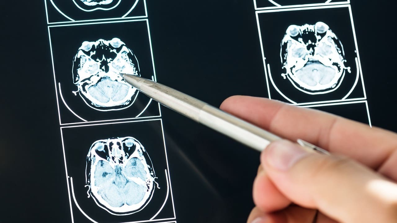
When we look at X-ray images of the human brain, we see the bones that cover it. The brain itself is not visible because X-rays can’t show soft tissues well.
The bones and skull shapes in X-ray images are very important. They tell us if the skull is okay or if there are problems like fractures or growth issues.
Radiologists check brain X-rays for many things. They look for signs of injury, infection, or other problems. They also check if the skull and its contents look normal.
Key features radiologists look for include:
X-ray imaging is great for showing skull bones, but MRI is better for soft tissues. MRI shows details of the brain, meninges, and other soft tissues.
Using X-rays with MRI or CT scans gives us a full picture. We can see both bones and soft tissues in the skull.
It’s important to check the safety of brain X-ray imaging, mainly for tumor risk. We need to look at the latest research and what it means for patient care.
Recent studies have looked into if X-rays can cause brain tumors. Some studies found a possible link, but others didn’t. A big review of studies found that the risk of brain tumors from X-rays is debated.
We must look closely at these studies. This includes their methods and how many people were studied. Most agree that the risk is low, but there might be some.
Research has also found a link between dental X-rays and non-cancerous tumors. Some studies say frequent dental X-rays might increase this risk. But, these findings need more study.
It’s also important to remember that dental X-rays and brain X-rays are different. The exposure and body areas are not the same.
Brain X-ray imaging needs careful thought for different patients. Kids are more sensitive to radiation because their bodies are growing. Adults have different risks based on their health and what they’re being checked for.
To show the balance, let’s look at a table:
| Patient Population | Benefits of Brain X-Ray | Potential Risks |
|---|---|---|
| Pediatric Patients | Quick diagnosis of skull injuries or abnormalities | Higher sensitivity to radiation, possible long-term risks |
| Adult Patients | Effective diagnosis of various brain conditions | Radiation exposure, possible risks for certain conditions |
| Elderly Patients | Helpful in diagnosing age-related brain conditions | Potential risks due to comorbidities, radiation exposure |
The table shows that benefits and risks vary by patient group. Healthcare providers can make better choices by considering these factors.
In conclusion, brain X-ray imaging has valid safety concerns and risks. But, its benefits are clear. By keeping up with research and weighing risks and benefits for each patient, we can offer top care while reducing risks.
In some medical cases, a brain X-ray is the best choice for checking brain injuries and issues. We look at when brain X-ray imaging is the best diagnostic tool.
In emergency cases, like traumatic brain injuries, a brain X-ray is often the first step. CT scans are used for kids who are very sick or have many injuries. But, X-rays are quick and effective for checking injury severity.
The benefits of using brain X-ray in emergencies include:
Brain X-ray imaging is cheaper than MRI or CT scans. This makes it a good choice for quick diagnosis without needing detailed images.
Doctors say, “Brain X-ray imaging is very cost-effective, which is key in places where advanced imaging is hard to get.” This shows how important brain X-ray is worldwide.
Brain X-ray imaging is easy to find in most healthcare places. This makes it a key tool in both rich and poor countries. Its availability and low cost make it a must-have in global health.
We find that brain X-ray is a key tool in many medical situations, like emergencies. Its low cost and easy access make it a big help in health care around the world.
Getting ready for your brain X-ray? We want to make sure you know what to expect. A brain X-ray is a test that doctors use to check your brain and skull’s health.
Before you go, you might need to take off any metal things like jewelry or glasses. When it’s time for the X-ray, a tech will help you get into the right spot. Afterward, you can go back to your day as usual.
It’s key to talk to your doctor about any worries you have. You might ask, “What will the X-ray show?” or “Do I need to do anything special?” Also, “How long will it take to get the results?”
Your doctor will go over the X-ray results with you. The images can show details about your skull, any breaks, or signs of other issues. It’s a good idea to ask your doctor to explain it in simple terms.
X-ray imaging of the brain is key in medical diagnostics. It has evolved to be more important, thanks to advances in CT technology. These improvements have made diagnoses more accurate.
Brain X-rays show important details about skull injuries and abnormalities. They might not show soft tissue well. But, with CT scans, doctors can now diagnose conditions like stroke and trauma more accurately.
As technology gets better, X-ray imaging will keep playing a big role in brain health. Knowing its strengths and weaknesses helps patients and doctors make better choices about tests.
A brain X-ray, also known as a skull X-ray, is a non-invasive test. It uses X-rays to show images of the brain and skull. We use it to check for skull injuries, detect changes, and diagnose conditions like fractures.
Traditional brain X-rays can’t see soft tissues well. We often need CT scans or MRI for a detailed brain view.
Modern brain X-ray technologies, like CT scans, give more detailed images. They are very accurate, helping to spot conditions like stroke and trauma.
The average dose for a brain X-ray is about 1.05 millisieverts. We follow protocols to keep radiation low, ensuring safety.
Brain X-ray images show the skull’s outline and structures. Our radiologists look for injuries, fractures, and other issues. They compare these images with MRI for a full brain understanding.
Yes, there are safety concerns, like tumor risk. Research links X-rays to non-cancerous tumors. We weigh risks and benefits for each patient.
Brain X-ray is good for emergencies, like head trauma, because it’s quick and affordable. We also consider global healthcare needs when choosing imaging.
To prepare, follow your doctor’s instructions, like removing jewelry. During the X-ray, you’ll be positioned. After, you can usually go back to normal activities. We help you understand your results and what to ask your doctor.
A brain X-ray looks at the skull, while CT and MRI show more of the brain. We pick the best imaging based on your needs.
We take special care with pregnant patients or those with conditions. We weigh risks and use safer options when we can.
Subscribe to our e-newsletter to stay informed about the latest innovations in the world of health and exclusive offers!