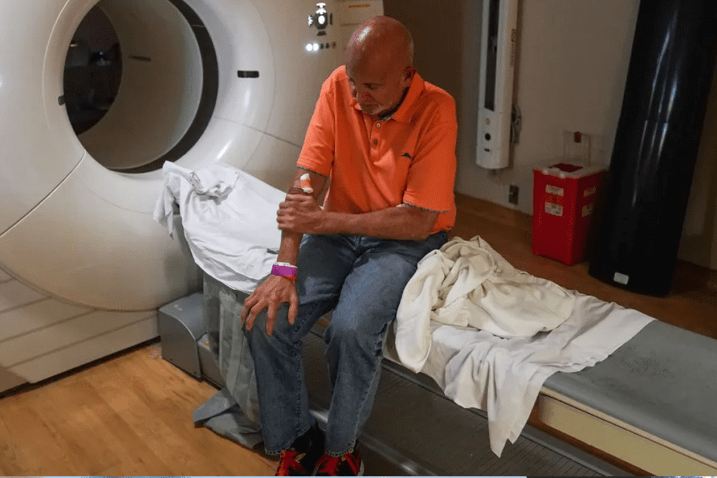
SPECT scans are a key tool in nuclear cardiology. Millions are done every year to check for different health issues.
They are mainly used to look at myocardial perfusion imaging. This helps find problems with blood flow to the heart. Such issues can cause serious heart problems.
SPECT scans are not just for the heart. They also help diagnose other conditions. This includes seizure disorders and some types of dementia. It shows how useful they are in medical tests.
Key Takeaways
- SPECT scans are used to diagnose a range of medical conditions.
- They are very helpful in checking heart blood flow issues.
- SPECT scans help in evaluating cardiac perfusion.
- These scans are also applied in diagnosing seizure disorders.
- Certain types of dementia can be diagnosed using SPECT scans.
The Science Behind SPECT Scan Technology

SPECT scans use nuclear medicine to see inside the body. They use tiny amounts of radioactive tracers. This technology helps doctors understand the body’s inner workings.
Basic Principles of Nuclear Medicine Imaging
Nuclear medicine uses tiny amounts of radioactive materials. These materials, or radionuclides, are given to the body to find and treat diseases. They emit gamma rays that the SPECT scanner catches.
First, a radiopharmaceutical is given to the body. It goes to the area it’s meant for. Then, as it decays, it sends out gamma rays. The SPECT scanner picks up these rays and turns them into images.
How SPECT Creates Three-Dimensional Images
SPECT makes 3D images by moving a camera around the patient. It takes data from many angles. Then, special algorithms turn this data into detailed 3D pictures.
These images show the body’s inside parts clearly. They help doctors find and fix many health problems. In cardiac imaging and cardiovascular imaging, SPECT is key. It shows how the heart works and spots issues like heart disease.
SPECT vs. Other Diagnostic Imaging Modalities
SPECT imaging is special because it shows how the body works. It gives us information about the body’s functions. This is different from other imaging methods that just show what the body looks like.
Comparing SPECT with CT and MRI
CT and MRI are two common imaging tools. CT scans give detailed pictures of the body’s inside. They are great for finding injuries and some tumors.
MRI shows soft tissues in high detail. It’s best for checking the brain, spinal cord, and muscles.
SPECT doesn’t show as much detail as CT or MRI. But, it tells us about how the body works. For example, in heart imaging, SPECT can show myocardial blood flow. This helps diagnose heart disease, like with a nuclear stress test.
Advantages of Functional vs. Structural Imaging
Functional imaging, like SPECT, shows how the body works. This is different from CT and MRI, which show what the body looks like. SPECT’s information is key for diagnosing and treating many conditions, like heart problems.
In some cases, SPECT’s functional info is more important than CT or MRI’s structural details. For example, SPECT can show how much of the heart is affected by disease. This helps doctors decide if surgery is needed.
When SPECT is Preferred Over Other Techniques
SPECT is best when we need to know how the body works. For heart disease, a nuclear stress test with SPECT is very helpful. It shows how the heart works under stress, helping find problems and predict future heart issues.
Compared to cardiac PET scan, SPECT is more common and established for some uses. But, PET might be better in some cases because it’s more sensitive and detailed.
Choosing between SPECT and other imaging depends on the situation and what’s available. Knowing what each can do is important for the best care.
Cardiac Perfusion: Primary Application of SPECT Imaging

Cardiac perfusion imaging through SPECT is a key tool for diagnosing heart issues. It shows how blood flows through the heart. This is very important for spotting coronary artery disease, a big cause of heart problems and death.
Myocardial Perfusion Imaging Explained
Myocardial perfusion imaging (MPI) is a test that checks the heart’s blood flow. It uses a tiny amount of radioactive tracer. This tracer shows how well blood flows to the heart muscle.
SPECT imaging then takes pictures of where the tracer goes. This helps doctors see if some parts of the heart don’t get enough blood.
Key aspects of MPI include:
- Assessment of coronary artery disease severity
- Identification of ischemia and infarction
- Guidance for revascularization decisions
Detecting Coronary Artery Disease
SPECT MPI is great at finding coronary artery disease (CAD). It shows how well the heart muscle gets blood. If some areas don’t get enough blood, it might mean there’s a blockage or narrowing in the arteries.
Quantifying Blood Flow to Heart Muscle
Knowing how much blood the heart muscle gets is very important. SPECT imaging can measure this. It helps doctors understand how severe CAD is and what treatment might be best.
| Parameter | Description | Clinical Significance |
| Myocardial Blood Flow (MBF) | Quantifies the blood flow to the heart muscle | Assesses the severity of CAD |
| Myocardial Flow Reserve (MFR) | Measures the capacity of the heart to increase blood flow | Predicts outcomes and guides treatment |
Cardiovascular Diseases Diagnosed Through SPECT
Cardiovascular diseases are a major cause of illness and death worldwide. SPECT scans are a key tool in diagnosing these diseases. They help doctors check how well the heart is working and find damaged areas.
Assessment of Cardiac Function and Ejection Fraction
SPECT imaging helps doctors measure the heart’s function. It calculates the ejection fraction (EF), which shows how well the heart pumps blood. A normal EF is between 55% and 70%. If it’s lower, the heart might not be working right.
Identifying Ischemia and Infarction
SPECT scans are great at finding problems with blood flow in the heart. They use special drugs that show up in healthy heart tissue. This helps doctors see where blood flow is low or where tissue is dead.
SPECT myocardial perfusion imaging is a cornerstone in the diagnosis and management of coronary artery disease, providing critical information on the presence and extent of ischemia.
Viability Assessment for Revascularization Planning
SPECT imaging is also used to see if heart tissue can recover. It helps doctors decide if a heart procedure is needed. This helps choose the best treatment for each patient.
| Condition | SPECT Imaging Findings | Clinical Implication |
| Ischemia | Reduced radiopharmaceutical uptake in stress images | Indicates coronary artery disease; may benefit from revascularization |
| Infarction | Permanent defect on both rest and stress images | Indicates scar tissue; management focuses on preventing further damage |
| Viable Myocardium | Uptake on rest or viability imaging | Potential benefit from revascularization |
Nuclear Stress Testing Protocols and Applications
Nuclear stress testing has changed cardiology. It shows how myocardial blood flow and heart function change under stress.
This test uses SPECT imaging to check the heart’s function under stress. It’s often done through exercise or medicine. It helps find coronary artery disease and guides treatment.
Rest vs. Stress SPECT Procedures
Nuclear stress testing has two SPECT scans: one at rest and one under stress. The resting scan shows the heart’s baseline. The stress scan shows how it works under stress.
By comparing these images, doctors can spot areas with less blood flow. This might mean coronary artery disease or other heart problems.
The main difference is how the scans are done. The resting scan comes first, then the stress scan. Stress can be from exercise or medicine that simulates exercise.
Exercise vs. Pharmacological Stress Methods
There are two ways to stress the heart during testing: exercise stress and pharmacological stress. Exercise stress uses physical activity to raise heart rate and blood flow. Pharmacological stress uses medicine to do the same without exercise.
Exercise stress testing is best for those who can exercise. It gives a more natural look at heart function. But, for those who can’t exercise, pharmacological stress is a good option.
The choice between exercise and medicine stress depends on the patient’s health and ability. Both methods help assess heart function under stress.
Neurodegenerative Disorders and SPECT Imaging
SPECT imaging is key in diagnosing neurodegenerative disorders. It shows how the brain works, helping to spot and tell apart different types of dementia.
Frontotemporal Dementia Identification
Frontotemporal dementia (FTD) damages the brain’s frontal and temporal lobes. SPECT imaging spots FTD by showing less blood flow in these areas. This is different from other dementias like Alzheimer’s, making diagnosis more precise.
Key features of FTD on SPECT imaging include:
- Reduced perfusion in the frontal and temporal lobes
- Asymmetric involvement, often more pronounced on one side
- Relative preservation of parietal and occipital lobe perfusion
Lewy Body Dementia Characteristics
Lewy body dementia (LBD) is another common dementia type. SPECT imaging in LBD shows less blood flow to the occipital lobe. This helps tell it apart from Alzheimer’s. LBD also shows ups and downs in blood flow, matching the patient’s symptoms.
The diagnostic utility of SPECT in LBD lies in its ability to:
- Demonstrate occipital lobe hypoperfusion
- Show relative preservation of medial temporal lobe structures
- Support the diagnosis when combined with clinical criteria
Vascular Dementia Assessment
Vascular dementia comes from reduced blood flow to the brain, often from small strokes. SPECT imaging shows where the brain is damaged by showing areas with less blood flow. This is key for managing risks and planning rehabilitation.
SPECT findings in vascular dementia may include:
- Multiple areas of reduced perfusion corresponding to vascular territories
- Perfusion defects that may be wedge-shaped or irregular
- Variable involvement of cortical and subcortical structures
SPECT in Epilepsy and Seizure Disorders
SPECT imaging is key in diagnosing epilepsy. It shows brain activity in detail. Finding where seizures start is vital for treatment.
Ictal vs. Interictal SPECT Studies
SPECT imaging can be done during or between seizures. Ictal SPECT captures brain activity during a seizure. It’s great for finding where seizures start.
Interictal SPECT is done between seizures. It shows the brain’s normal activity. Comparing these images helps pinpoint seizure locations better.
SISCOM Technique for Enhanced Seizure Localization
The SISCOM technique is advanced. It subtracts images taken between seizures from those during seizures. Then, it matches the result with MRI. This method clearly shows where seizures happen.
“SISCOM has changed epilepsy surgery,” studies say. It helps find the seizure cause more accurately. It’s very helpful for complex cases.
The use of SISCOM in epilepsy diagnosis is a big step forward. It helps doctors understand seizures better and treat them more effectively.
Using SPECT and SISCOM improves diagnosis and treatment for epilepsy patients.
Neuropsychiatric Conditions Evaluated with SPECT
SPECT helps us understand brain function and disease. It’s a key tool in neuropsychiatry. It lets doctors check mental health and brain function.
ADHD Brain Function Patterns
ADHD shows unique brain activity patterns. SPECT imaging shows these patterns. It finds less activity in areas like the prefrontal cortex, key for focus and control.
Depression and Anxiety Disorders
SPECT also looks at depression and anxiety. These conditions change brain blood flow, mainly in the limbic system. This helps doctors understand the brain’s role in these disorders.
| Condition | Brain Region Affected | SPECT Findings |
| ADHD | Prefrontal Cortex | Decreased perfusion |
| Depression | Limbic System | Altered perfusion patterns |
| Anxiety Disorders | Amygdala | Increased activity |
PTSD and Trauma-Related Brain Changes
PTSD can also be studied with SPECT. People with PTSD have brain activity changes. These changes are in areas for emotions and memory. SPECT shows these changes, helping in diagnosis and treatment.
With SPECT, doctors learn more about brain disorders. This knowledge helps in creating better treatments. It improves how patients are cared for.
DaTscan: Specialized SPECT for Movement Disorders
Dopamine transporter imaging with DaTscan is key for diagnosing Parkinson’s disease and other movement disorders.
DaTscan uses a special radiopharmaceutical that binds to dopamine transporters in the striatum. This lets doctors check dopamine transporter density. It’s very helpful in diagnosing and differentiating various movement disorders.
Dopamine Transporter Imaging Principles
DaTscan works by using a specific radiopharmaceutical that targets dopamine transporters. These transporters are on the presynaptic terminals of dopaminergic neurons. They play a big role in regulating dopamine levels in the synaptic cleft.
Key aspects of dopamine transporter imaging include:
- Specific binding to dopamine transporters
- Assessment of dopamine transporter density
- Use in SPECT imaging to provide functional information about the dopaminergic system
Differentiating Parkinson’s Disease from Look-alikes
Diagnosing Parkinson’s disease can be tricky because it looks like other conditions. DaTscan is very helpful in this area.
| Condition | DaTscan Pattern | Clinical Implication |
| Parkinson’s Disease | Reduced dopamine transporter binding, often asymmetric | Supports the diagnosis of Parkinson’s Disease |
| Essential Tremor | Normal dopamine transporter binding | Helps rule out Parkinson’s Disease |
| Drug-induced Parkinsonism | Normal dopamine transporter binding | Distinguishes from Parkinson’s Disease |
Evaluating Parkinsonian Syndromes
DaTscan is not just for Parkinson’s disease. It’s also great for checking other parkinsonian syndromes. The dopamine transporter binding pattern gives clues about the disease.
Using DaTscan in clinics has made diagnosing better. It helps doctors see how much dopamine transporter there is. This helps them tell different conditions apart and decide on the best treatment.
Bone SPECT Applications in Orthopedics
In orthopedics, Bone SPECT is key for checking complex bone and joint problems. It’s great for tough cases that other images can’t solve.
Diagnosing Occult Fractures and Stress Injuries
Bone SPECT is top-notch for finding hidden fractures and stress injuries. These are hard to spot on regular X-rays or some other scans. It shows how bones are working, helping find active areas that might be broken or stressed.
A study in a Journa showed Bone SPECT’s power. It found fractures that other images missed.
Bone SPECT has emerged as a valuable tool in the diagnosis of occult fractures, showing it’s good at finding fractures not seen on other images.
Evaluating Complex Regional Pain Syndrome
CRPS is hard to diagnose, needing both doctor checks and scans. Bone SPECT helps by showing how bones act in CRPS.
| Characteristics | Bone SPECT Findings |
| Increased bone metabolism | More tracer uptake in the affected limb |
| Regional osteoporosis | Less bone density all over |
Assessing Joint Prosthesis Complications
Bone SPECT is also good for checking on joint prostheses. It spots problems like loosening or infection early. This helps doctors act fast.
A study in a Journal showed Bone SPECT/CT is better than regular X-rays for finding loose prosthetics.
In summary, Bone SPECT is very useful in orthopedics. It helps find hidden fractures, check CRPS, and spot problems with joint prostheses. Its detailed insights make it a key tool for doctors.
Infection and Inflammation Detection with SPECT
SPECT is a key tool in nuclear medicine for spotting infections and inflammation. It helps doctors diagnose and treat many conditions. This gives them the insights they need to make the right treatment plans.
Osteomyelitis Diagnosis and Monitoring
Osteomyelitis, or bone infection, is hard to spot with regular imaging. But SPECT scans, with the right drugs, can find it and track how it’s doing. This helps doctors plan the best treatment, like surgery or antibiotics.
Key Benefits of SPECT in Osteomyelitis:
- Early detection of infection
- Assessment of infection extent
- Monitoring treatment response
Gallium-67 and Indium-111 for Infection Imaging
Gallium-67 and Indium-111 are drugs used in SPECT scans for finding infections. Gallium-67 goes to inflamed or infected areas, helping spot many types of infections. Indium-111, on the other hand, targets infections by gathering in infected tissues.
| Radiopharmaceutical | Application | Advantages |
| Gallium-67 | General infection imaging | Effective for various infections |
| Indium-111 | Specific infection localization | High sensitivity for infected tissues |
Inflammatory Bowel Disease Assessment
SPECT helps doctors check how severe inflammatory bowel disease (IBD) is. This info is key for managing IBD. It helps doctors decide on treatments and keep an eye on how the disease is doing.
SPECT’s role in finding infections and inflammation shows its importance in medicine. It gives doctors detailed info on diseases. This helps in giving better care to patients.
Endocrine System Disorders Diagnosed by SPECT
SPECT imaging is key in checking the endocrine system. It helps us see how different glands work. The endocrine system has glands that make hormones. These hormones control growth, metabolism, and more.
Endocrine system problems can be hard to spot. But SPECT scans help find and fix these issues.
Parathyroid Adenoma Localization
Parathyroid adenomas are small tumors that can make too much hormone. SPECT scans find these tumors. This helps doctors plan better surgeries.
- Improved surgical planning
- Enhanced accuracy in adenoma localization
- Reduced risk of complications during surgery
Thyroid Nodule Evaluation and Cancer Screening
SPECT imaging checks thyroid nodules. It tells doctors if a nodule might be cancer. SPECT shows how well a nodule works.
- Assessment of nodule function
- Evaluation of nodule metabolism
- Guidance for fine-needle aspiration biopsy
Adrenal Gland Imaging Applications
The adrenal glands make hormones for our body. SPECT imaging looks at adrenal gland problems. This includes Cushing’s syndrome and pheochromocytoma.
| Adrenal Gland Disorder | SPECT Imaging Application |
| Cushing’s syndrome | Assessment of adrenal gland hyperfunction |
| Pheochromocytoma | Localization of adrenal gland tumors |
Oncological Applications of SPECT Imaging
SPECT imaging is key in fighting cancer. It helps doctors find and treat tumors better. It shows how tumors grow and spread, helping plan treatments.
Tumor Detection and Characterization
SPECT scans find and study tumors. They use special medicines to spot cancer cells. This helps tell if a tumor is bad or not.
Special medicines make tumors show up clearer on SPECT scans. This means doctors can see cancer cells better.
Sentinel Node Mapping in Breast Cancer and Melanoma
SPECT is great for finding the first lymph node cancer reaches. This is called the sentinel node. Knowing this helps doctors understand how far cancer has spread.
Using SPECT with CT scans makes finding this node even better. It gives doctors a clear picture of where to operate.
Neuroendocrine Tumor Imaging with MIBG
Neuroendocrine tumors come from special cells. SPECT with MIBG is good for finding these tumors. It works for pheochromocytomas and neuroblastomas.
MIBG goes into cells it should, showing tumors. This helps doctors find and treat these tumors better.
In short, SPECT imaging is very important in fighting cancer. It helps find tumors, map cancer spread, and find neuroendocrine tumors. Its role keeps growing, helping doctors fight cancer better.
Pulmonary Conditions Assessed with SPECT
SPECT imaging is a key tool in diagnosing lung diseases. It gives detailed information about lung function. This makes it great for checking different lung conditions.
Pulmonary Embolism Diagnosis
Pulmonary embolism is a serious issue where a blood clot blocks a lung artery. SPECT imaging is vital in diagnosing this condition. It’s very helpful for patients who can’t use other imaging methods.
The benefits of SPECT for diagnosing pulmonary embolism include:
- It’s very good at finding clots in the lungs.
- It’s great for patients who are very sick or can’t have other tests.
- It gives useful information for deciding on treatment.
Ventilation-Perfusion Studies for Lung Function
Ventilation-perfusion (V/Q) studies are a type of SPECT imaging. They check how well the lungs breathe and get blood. This is very useful for diagnosing lung function problems.
V/Q SPECT is good for:
- Finding pulmonary embolism by showing which parts of the lung are breathing but not getting blood.
- Checking lung function before surgery.
- Seeing how chronic lung diseases affect lung function.
Pre-operative Evaluation for Lung Resection
Before lung surgery, SPECT imaging helps check lung function. This helps plan the surgery and predict how well the lungs will work after.
The info from V/Q SPECT helps surgeons:
- Figure out how much lung tissue can be safely removed.
- Plan the surgery to keep as much lung function as possible.
- Tell patients what to expect and the possible risks.
Emerging Applications and Hybrid Imaging
SPECT technology is changing how we diagnose diseases. It’s now part of hybrid imaging, combining different imaging types. This gives us more detailed information for better diagnosis.
SPECT/CT Fusion Imaging Benefits
SPECT/CT fusion imaging is a big step forward. It mixes SPECT’s function info with CT’s anatomy. This combo gives us a clearer picture of diseases.
It helps doctors find and understand diseases better. This is key in cancer, heart, and infection cases.
Novel Radiopharmaceuticals in Development
New radiopharmaceuticals are being made for SPECT. These tracers target specific diseases, helping diagnose them earlier and more accurately. They bind to certain receptors or proteins, showing us specific disease processes.
These new agents will let us diagnose more conditions with SPECT. This will make SPECT even more useful in medicine.
Quantitative SPECT for Precision Medicine
Quantitative SPECT is a big step towards precision medicine. It lets us measure how much tracer is taken up. This helps us see how severe a disease is and how it’s changing.
Using quantitative SPECT in medicine will lead to better patient care. It helps tailor treatments to each patient’s needs.
Limitations, Risks, and Considerations
SPECT scans are useful for diagnosis but have some limits and risks. It’s important for healthcare providers and patients to understand these. This helps in making informed choices.
Radiation Safety and Exposure
SPECT scans use small amounts of radioactive tracers. This can expose patients to radiation. To reduce this risk, safety steps are taken. These include using the least amount of radioactive material and monitoring patients closely.
Patient Preparation and Contraindications
Getting ready for a SPECT scan is key for accurate results. Some conditions, like pregnancy or severe kidney disease, might not allow SPECT scans. Doctors must check if a patient is suitable before the scan.
Cost-Effectiveness and Insurance Coverage
The cost of SPECT scans is a big factor. They can give important diagnostic info but are pricey. Insurance coverage varies. Patients should check their insurance before getting a SPECT scan.
FAQ
What is a SPECT scan used for?
A SPECT scan is a test that uses nuclear medicine. It helps doctors diagnose and track many health issues. This includes heart disease, brain disorders, and some cancers.
How does SPECT scan technology work?
A SPECT scan detects gamma rays from a special drug injected into the body. A camera captures images from different angles. This creates a 3D picture of the body’s inner workings.
What is the difference between SPECT and other diagnostic imaging modalities like CT and MRI?
SPECT shows how tissues work, unlike CT and MRI which focus on body structure. SPECT looks at blood flow and metabolism. It’s great for checking on the body’s functions.
What is cardiac perfusion imaging, and how is it used to diagnose coronary artery disease?
Cardiac perfusion imaging is a SPECT scan for the heart. It checks blood flow to the heart muscle. It spots blockages or narrowings in the heart’s arteries.
Can SPECT scans be used to diagnose neurodegenerative disorders like Alzheimer’s disease?
Yes, SPECT scans can help find and track neurodegenerative diseases like Alzheimer’s. They look for brain activity patterns specific to these conditions.
How is SPECT used in epilepsy and seizure disorders?
SPECT helps find where seizures start in the brain. It uses special studies to pinpoint the seizure focus. This helps doctors treat epilepsy better.
What is DaTscan, and how is it used to diagnose movement disorders?
DaTscan is a SPECT scan for the brain. It looks at dopamine transporters. It helps diagnose Parkinson’s disease and other movement disorders.
Can SPECT scans be used to detect bone metastases or diagnose osteomyelitis?
Yes, SPECT scans can find bone metastases and osteomyelitis. They’re good for spotting hidden fractures and checking joint replacements.
How is SPECT used in oncological applications, such as tumor detection and sentinel node mapping?
SPECT helps find and study tumors in cancer. It’s also used to map sentinel nodes in breast cancer and melanoma. It can image neuroendocrine tumors too.
References
Maffeis, C., et al. (2023). Clinical application of myocardial perfusion SPECT in assessing coronary artery disease.


