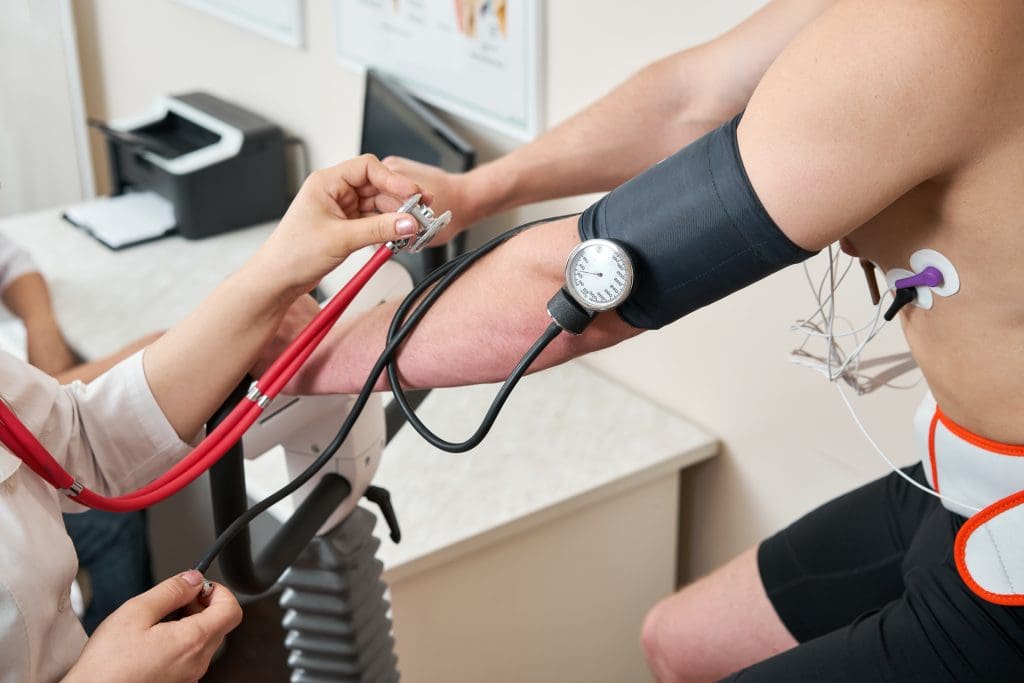
Cardiology is the medical specialty that focuses on heart health and cardiovascular diseases. It uses many methods and tools to check heart health.
Nuclear cardiology is a part of cardiology. It uses special imaging and radioactive materials to look at the heart. The American Society of Nuclear Cardiology says it’s key for finding and understanding heart diseases.
It’s important to know the difference between cardiology and nuclear cardiology. Cardiology gives a wide view of heart health. But nuclear cardiology gives a closer look at how the heart works and any problems it might have.

Cardiology is a key field in keeping our hearts healthy. It deals with finding, treating, and preventing heart and blood system problems.
Cardiology covers a wide range of tasks, from early prevention to advanced treatments. Cardiologists are experts who focus on heart diseases. They use many tools and methods to diagnose and treat.
They handle issues like heart blockages, failure, and irregular heartbeats. Cardiologists team up with other doctors to give full care.
To be a cardiologist, one needs a lot of education. This includes medical school, internal medicine residency, and cardiology fellowship.
To get certified, cardiologists pass the American Board of Internal Medicine (ABIM) test. They must keep learning and follow professional rules to stay certified.
Cardiologists use many tools to check heart health. These include:
These tools help cardiologists find and treat heart issues. This leads to better health for patients.
Nuclear cardiology is a big step forward in fighting heart diseases. It uses nuclear medicine to check how well the heart works. This field uses special substances and high-tech images to see the heart’s health.
Nuclear cardiology uses tiny amounts of radioactive tracers to see the heart. It uses SPECT and PET imaging to check the heart’s blood flow and health. This helps doctors find and fix heart problems.
Nuclear cardiology tests give deep insights into the heart. They help doctors spot heart issues, see how bad heart disease is, and check if treatments work.
Nuclear cardiology has grown a lot over time. It started with early nuclear medicine. New tech and tracers have made it better. Now, it’s a key part of heart disease care.
Nuclear cardiology is close to general nuclear medicine. But, it focuses only on the heart. It uses special methods for heart images, making it unique.
The Journal of Nuclear Cardiology helps experts in this field. It shares new research and rules to improve nuclear cardiology.
Cardiology and nuclear cardiology are related but different. They use different methods to diagnose and treat heart issues. The main differences are in how they test patients, the technology used, and the patient experience.
Cardiology often uses tests like ECGs, echocardiograms, and stress tests. Nuclear cardiology, on the other hand, uses radioactive tracers and advanced imaging like SPECT and PET. These help see the heart’s structure and function.
Nuclear cardiology gives detailed info on the heart’s blood flow and function. This is key for diagnosing coronary artery disease and checking heart muscle before surgery.
Cardiology and nuclear cardiology use different technologies. Cardiology has tools like ECG machines and echocardiography systems. Nuclear cardiology needs special equipment, like gamma cameras for SPECT and PET scanners.
Radioactive tracers in nuclear cardiology let doctors see how well the heart is working. This info isn’t available with traditional cardiology tests.
| Aspect | Cardiology | Nuclear Cardiology |
| Diagnostic Tools | ECG, Echocardiogram, Stress Test | SPECT, PET, Radioactive Tracers |
| Primary Focus | Heart Structure and Function | Myocardial Perfusion and Viability |
| Equipment | ECG Machines, Echocardiography Systems | Gamma Cameras, PET Scanners |
The patient experience varies between cardiology and nuclear cardiology. Nuclear cardiology tests use radioactive tracers. This means patients might need to prepare differently, like avoiding certain meds or fasting.
During the test, patients lie under the camera for a long time. This can be uncomfortable.
Even with these differences, both cardiology and nuclear cardiology are vital for heart disease diagnosis and management. The choice between them depends on the specific needs of the patient.
Nuclear cardiology uses different tests to check the heart’s health. Each test gives unique insights into how well the heart works.
Myocardial perfusion imaging is key in nuclear cardiology. It checks how well blood flows to the heart muscle. It finds areas where blood flow is low, which can mean heart disease.
SPECT (Single Photon Emission Computed Tomography) and PET (Positron Emission Tomography) are main methods. SPECT gives detailed images of blood flow. PET is more sensitive and great for finding heart disease.
Cardiac blood pool scans, or MUGA scans, check the heart’s pumping. A small radioactive tracer is injected into the blood. This lets doctors see how well the heart’s ventricles pump blood.
MUGA scans help monitor heart function in patients with heart issues or those getting chemotherapy. They measure how well the heart pumps and moves.
Cardiac PET metabolic imaging looks at the heart’s metabolic activity. It uses special tracers to see how well heart tissue works. This helps manage heart failure and coronary artery disease.
This method is great for finding heart tissue that’s not working but can be saved. It helps decide if revascularization is needed.
| Test Type | Description | Primary Use |
| Myocardial Perfusion Imaging (SPECT) | Assesses blood flow to the heart muscle using SPECT technology | Diagnosing coronary artery disease |
| Myocardial Perfusion Imaging (PET) | Evaluates blood flow and metabolism using PET technology | Detecting coronary artery disease with high sensitivity |
| Cardiac Blood Pool Scans (MUGA) | Evaluates the heart’s pumping function | Monitoring heart function in chemotherapy patients or those with heart conditions |
| Cardiac PET Metabolic Imaging | Assesses the metabolic activity of the heart | Managing heart failure and assessing myocardial viability |
Nuclear cardiology offers detailed heart images. It’s used for checking coronary artery disease, heart failure, and pre-operative risks.
Nuclear cardiology is key in diagnosing coronary artery disease. Myocardial perfusion imaging (MPI) shows ischemia and infarction areas.
In heart failure, nuclear cardiology checks left ventricular function and viability. This helps choose the best treatment.
Nuclear cardiology checks cardiac risk before surgery. It spots high-risk patients for needed precautions.
After surgeries like revascularization, nuclear cardiology checks treatment success. It looks for any leftover ischemia.
| Clinical Application | Description | Nuclear Cardiology Technique |
| Coronary Artery Disease Assessment | Diagnosing and managing CAD | Myocardial Perfusion Imaging (MPI) |
| Heart Failure Evaluation | Evaluating left ventricular function and myocardial viability | Cardiac Blood Pool Scans (MUGA) |
| Pre-operative Risk Assessment | Assessing cardiac risk before non-cardiac surgery | Myocardial Perfusion Imaging (MPI) |
Getting ready for nuclear cardiology tests is important. It helps reduce stress and makes the process smoother. Knowing what to expect before, during, and after can improve your experience.
There are steps to take before the test to get accurate results. These include:
It’s key to follow the exact instructions from your healthcare provider or the testing center. These can change based on the test type.
During the test, you’ll lie on a table while images are taken. A radioactive tracer might be used to see how your heart works and blood flows.
You might feel a bit uncomfortable because you have to stay very quiet for a while. Or, if it’s a stress test, you might feel stressed. But, most people find it okay, and the staff will help and watch over you.
After the test, you can usually go back to your normal activities. The tracer used will lose its radioactivity and leave your body over time.
As the American Society of Nuclear Cardiology says, “You’ll get your test results in a few days. A cardiologist or nuclear medicine specialist will explain them to you.”
“Nuclear cardiology tests give important info about your heart. They help find and manage heart disease well.” –
American Society of Nuclear Cardiology
Make sure to talk to your healthcare provider about the results. They’ll tell you what to do next based on what the test showed.
Nuclear cardiology needs special training and certification. This ensures professionals use its tools well. It’s a field that mixes cardiology and nuclear medicine.
The certification process for nuclear cardiology is tough. It makes sure professionals know their stuff. The American Society of Nuclear Cardiology (ASNC) offers help and programs for certification.
To get certified, you need to finish some education, pass a test, and have enough experience. The test checks your knowledge in radiation safety, nuclear cardiology, and reading images.
Getting certified shows a nuclear cardiologist’s skill and dedication to good patient care.
Nuclear cardiologists must keep up with new discoveries. They do this by taking more classes, going to workshops, and reading new research. The ASNC has many educational tools, like online courses and conferences.
They also need to:
To keep certification, nuclear cardiologists must keep learning. The ASNC has rules for this, like getting a certain number of education credits every few years.
| Certification Requirement | Description | Frequency |
| Continuing Education Credits | Get a set number of credits in nuclear cardiology | Every 3 years |
| Professional Development | Take part in workshops, conferences, and online courses | Ongoing |
| Certification Renewal | Renew certification through the ASNC | Every 3-5 years |
The ASNC says, “Keeping up with new tech and methods is key for nuclear cardiologists.” This shows how important it is to keep learning for the best patient care.
Advancements in nuclear cardiology come from professional groups and academic sources. These groups are key in pushing research, education, and clinical practice forward.
The American Society of Nuclear Cardiology (ASNC) leads in nuclear cardiology. It offers guidelines, educational tools, and networking chances for experts.
The Journal of Nuclear Cardiology is a major academic source. It publishes new research and reviews on nuclear cardiology. It’s a key place for sharing knowledge and moving the field forward.
Other groups also help in nuclear cardiology. These include:
Both patients and professionals gain from these organizations. For patients, there are:
For professionals, there are:
By using these resources, both patients and professionals can keep up with nuclear cardiology’s latest.
Nuclear cardiology is a key part of cardiology, helping to diagnose and manage heart diseases. It uses special tools that work alongside traditional methods. But, it also has its own set of challenges, like the risk of radiation exposure.
Nuclear cardiology has many diagnostic advantages. It can check how well the heart muscle is working and spot coronary artery disease. It also helps see if treatments are working.
Tests like Single Photon Emission Computed Tomography (SPECT) and Positron Emission Tomography (PET) give detailed heart images. This helps doctors diagnose and treat complex heart conditions.
These tests can find coronary artery disease early. This means doctors can start treatments sooner, improving patient outcomes. They also help figure out if heart muscle can recover, guiding treatment for heart failure or after heart surgeries.
A big challenge of nuclear cardiology is radiation exposure. The tests use radiopharmaceuticals, which expose patients to ionizing radiation. This can increase the risk of cancer and genetic changes.
But, the benefits of these tests often outweigh the risks. This is true when they are used carefully and with the right amount of radiation.
To lower radiation exposure, new, more efficient radiopharmaceuticals are being made. Also, dose reduction strategies are being used. For example, stress-only SPECT tests can cut down radiation doses. New imaging tech, like CZT cameras, also helps by making images clearer with less radiation.
The cost and accessibility of nuclear cardiology tests are key issues. These tests are pricier than some other heart imaging, like echocardiography. This is because they need special equipment and radiopharmaceuticals.
Not everyone can get these tests because they are not available everywhere. But, more places are getting the needed equipment. This makes these tests more accessible and affordable for more people.
The future of nuclear cardiology is bright, thanks to new tech and research. Several areas are leading the way in this field.
Nuclear cardiology is getting better with new imaging tech. This includes new medicines and better scanners. These advancements make tests more accurate and useful.
Research in nuclear cardiology is pushing the field forward. It’s focused on new uses and better methods. Some key areas include:
The future of nuclear cardiology is in combining it with other heart imaging. This mix gives a deeper look at the heart’s health.
By joining forces with other imaging, nuclear cardiology can help patients more. We can expect more breakthroughs in hybrid imaging and personalized care.
Nuclear cardiology is a key medical field that has changed how we diagnose and treat heart issues. It’s important for doctors and patients to know the difference between cardiology and nuclear cardiology.
Nuclear cardiology has grown a lot, using new technologies and methods for precise diagnoses and treatments. It’s a part of cardiology that uses nuclear medicine to check heart function and find heart diseases.
The role of nuclear cardiology in today’s medicine is huge. It helps manage heart disease, heart failure, and other heart problems. With nuclear cardiology, doctors can make better choices, help patients more, and improve care quality.
As nuclear cardiology keeps growing, it’s vital to keep up with new findings and advancements in cardiology and nuclear medicine.
Nuclear cardiology uses tiny amounts of radioactive tracers to help diagnose and manage heart disease. It’s a part of cardiology and nuclear medicine.
Cardiology is a wide field that deals with heart and blood vessel disorders. Nuclear cardiology is a part of it. It focuses on using nuclear techniques for heart disease diagnosis and management.
Cardiology uses many tools like electrocardiograms and echocardiograms. Stress tests and cardiac catheterization are also common. Nuclear cardiology tests, like myocardial perfusion imaging, are used too.
Myocardial perfusion imaging uses radioactive tracers to check blood flow to the heart. It helps find coronary artery disease and check treatment success.
Preparing for a test varies by type. Usually, avoid certain foods and drinks beforehand. Wear comfy clothes. Tell your doctor about any meds you’re on.
Nuclear cardiology helps find heart disease early and accurately. It shows disease severity and treatment success. It’s a key tool in heart care.
It uses radiation, which some find concerning. Tests can be pricey and not always available everywhere.
The American Society of Nuclear Cardiology promotes nuclear cardiology in heart disease diagnosis and management. It offers education and supports its use in practice.
The Journal of Nuclear Cardiology publishes research and reviews on nuclear cardiology. It’s a key resource for professionals.
Certification comes from the Certification Board of Nuclear Cardiology. You need education, training, and pass an exam to become certified.
Nuclear cardiology’s future includes new technologies and techniques. Research aims to improve test accuracy and effectiveness.
Subscribe to our e-newsletter to stay informed about the latest innovations in the world of health and exclusive offers!