
Getting a correct diagnosis for groin hernias is key for good treatment. Femoral hernias are rare, making up about 3 to 5 percent of all groin hernias. They can be tricky to tell apart from inguinal hernias on CT scans.
Femoral hernias are more common in women and have a higher risk of serious problems like strangulation. StatPearls says they happen less often than inguinal hernias. It’s important to get a correct diagnosis with CT scans for the right treatment.
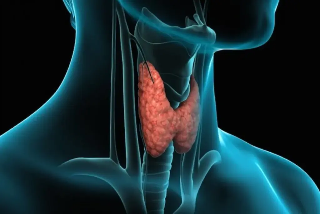
Liv Hospital is known for its patient-focused approach in diagnosing and treating hernias. Knowing how to spot the difference between femoral and inguinal hernias on CT scans is vital for doctors and patients.
Key Takeaways
- Accurate diagnosis of groin hernias is critical for effective treatment.
- Femoral hernias are more common in females and have a higher risk of complications.
- CT scans play a vital role in distinguishing between femoral and inguinal hernias.
- Liv Hospital offers expertise in radiology with a patient-centered approach.
- Understanding CT scan differences is essential for clinicians and patients.
Groin Hernia Fundamentals: Epidemiology and Clinical Significance

Knowing about groin hernias is key for radiologists. They need to tell femoral from inguinal hernias. These hernias can really affect a person’s life, so quick and right diagnosis is vital.
Prevalence of Femoral vs Inguinal Hernias
Many people get groin hernias over their lifetime. Men have a 27% to 43% chance, and women have a 3% to 6% chance. Inguinal hernias are more common, but femoral hernias can lead to serious problems.
Prevalence Rates:
| Hernia Type | Male Prevalence | Female Prevalence |
| Inguinal Hernia | 27%-43% | 3%-6% |
| Femoral Hernia | Rare | More common than in males |
Gender Distribution and Risk Factors
Men and women get groin hernias differently. Femoral hernias are more common in women, while men get inguinal hernias more often. Age, family history, and high abdominal pressure are risk factors.
Complication Rates and Clinical Importance
Groin hernias can cause serious problems like incarceration and strangulation. These need emergency surgery. Accurate diagnosis is very important for treatment and patient care.
Complication Rates:
- Femoral hernias have a higher risk of strangulation.
- Inguinal hernias are more likely to be incarcerated.
- Prompt diagnosis is critical to prevent complications.
Anatomical Foundations of the Inguinal and Femoral Regions

It’s key for radiologists to know the anatomy of the inguinal and femoral regions. These areas are prone to hernias, making diagnosis tricky.
Critical Anatomical Landmarks for Radiologists
Radiologists need to know several key landmarks when looking at CT scans. These include:
- The inguinal ligament, a dividing line between the inguinal and femoral areas.
- The pubic tubercle, a bony landmark for hernia location.
- The femoral vein, a major blood vessel related to femoral hernias.
The Inguinal Ligament as a Dividing Structure
The inguinal ligament connects the anterior superior iliac spine to the pubic tubercle. It separates the inguinal and femoral regions. Knowing where a hernia is in relation to this ligament is key for diagnosis.
Vascular Relationships in the Groin
The groin has many blood vessels, with the femoral vein being a major one. Femoral hernias are often near this vein. On a femoral hernia ct scan, the hernia sac is usually medial to the femoral vein.
Important vascular relationships include:
- The femoral artery and vein, key in the femoral region.
- The inferior epigastric vessels, important for inguinal hernias on ct.
Knowing these anatomical and vascular details helps radiologists make accurate diagnoses from ct of inguinal hernia or inguinal hernia on ct scans.
CT Imaging Techniques for Optimal Hernia Evaluation
Getting the best CT scans is key to telling femoral and inguinal hernias apart. The quality of the scan and the imaging techniques used are very important.
Standard Protocol Parameters
Using the same CT scan settings is important for clear images. This helps doctors accurately check for hernias. Key settings include:
- Slice thickness: Thin slices (≤1 mm) for detailed views
- Pitch: Adjusted for the best image quality
- kVp and mAs: Set based on patient size and scan area
- Reconstruction interval: Overlapping for better multiplanar views
These standard settings help make sure CT scans are clear and consistent. This is vital for diagnosing hernias.
| Parameter | Recommended Setting | Benefit |
| Slice Thickness | ≤1 mm | Detailed view of hernia anatomy |
| Pitch | Adjusted for image quality | Best image quality for accurate diagnosis |
| kVp and mAs | Tailored to patient size | Right radiation dose and image quality |
Coronal-Oblique Reconstruction Benefits
Coronal-oblique reconstructions are great for looking at groin hernias. They give a clearer view of the hernia and its surroundings.
Benefits include:
- Clearer view of the hernia sac and its contents
- Better understanding of hernia size and location
- How the hernia relates to important structures like the inguinal ligament
Contrast Enhancement Considerations
Using contrast in CT scans can help diagnose hernias better. But, it’s important to think about the risks.
Considerations for contrast enhancement:
- Clearer view of blood vessels
- Clearer view of the hernia sac and its contents
- Risks like allergic reactions or kidney damage
By carefully thinking about these points, doctors can improve CT scans for hernia checks. This leads to more accurate diagnoses and better care for patients.
Femoral Hernia CT Scan: Diagnostic Hallmarks
Diagnosing femoral hernias on CT scans is key for good treatment plans. These hernias have special signs on scans that help doctors a lot.
Characteristic Location and Boundaries
Femoral hernias sit below the inguinal ligament, unlike inguinal hernias. On CT scans, this means they are below the ligament and next to the femoral vessels. The exact spot is very important for treatment and avoiding problems.
The Venous Compression Sign
One big sign of femoral hernias on CT scans is how they press on nearby veins, like the femoral vein. The venous compression sign is a big clue. It shows up as a smaller vein and a bigger vein up ahead.
“The compression of the femoral vein by a femoral hernia is a critical radiological finding that aids in diagnosis.”
Funnel-Shaped Neck Morphology
Femoral hernias have a funnel-shaped neck on CT scans. This shape is narrow at the top and wider at the bottom. It happens because the hernia goes through a narrow space.
To sum up, femoral hernias on CT scans have a few key signs. These include their location, the venous compression sign, and the funnel-shaped neck. Spotting these signs helps doctors diagnose femoral hernias correctly.
Inguinal Hernia on CT Scan: Key Imaging Features
CT scans are key for spotting inguinal hernias and what’s around them. Radiologists look for certain signs that show these hernias apart from others.
Medial Position to Pubic Tubercle
Inguinal hernias show up on CT scans in a specific spot. They are usually medial to the pubic tubercle. This spot is important for telling inguinal hernias from other groin hernias.
The Lateral Crescent Sign
The lateral crescent sign is another clue for inguinal hernias on CT scans. It looks like a crescent shape in the hernia sac. Seeing this sign helps confirm an inguinal hernia diagnosis.
Extended Sac Characteristics
Inguinal hernias have an extended sac seen on CT scans. The sac’s size, shape, and what’s inside give clues for diagnosis and planning before surgery. It might hold bowel loops, omentum, or other things.
| Feature | Description | Clinical Significance |
| Position relative to pubic tubercle | Medial to pubic tubercle | Aids in differentiating inguinal hernias |
| Lateral Crescent Sign | Crescent-shaped portion of hernia sac | Confirms diagnosis of inguinal hernia |
| Extended Sac Characteristics | Size, shape, and content of hernia sac | Provides information for diagnosis and preoperative planning |
By looking closely at these signs on CT scans, radiologists can accurately spot inguinal hernias. This helps a lot with planning for surgery.
Femoral Versus Inguinal Hernia: The 7 Critical CT Differences
It’s important to know the differences between femoral and inguinal hernias for the right treatment. CT scans help spot these differences. This is key for doctors to make good decisions.
Difference #1: Relationship to the Inguinal Ligament
Femoral hernias are below the inguinal ligament. Inguinal hernias are above it. This is a main difference seen on CT scans.
Difference #2: Position Relative to Pubic Tubercle
Inguinal hernias are near the pubic tubercle. Femoral hernias are more to the side and below it.
Difference #3: Hernia Neck Configuration
Femoral hernias have a funnel-shaped neck. Inguinal hernias have a cylindrical or saccular shape.
Difference #4: Vascular Compression Patterns
Femoral hernias often press on the femoral vein. This is rare in inguinal hernias. This can cause specific symptoms and is a key sign.
| Characteristic | Femoral Hernia | Inguinal Hernia |
| Relation to Inguinal Ligament | Below the inguinal ligament | Above the inguinal ligament |
| Position Relative to Pubic Tubercle | Laterally and inferiorly | Medially |
| Hernia Neck Configuration | Funnel-shaped | Cylindrical or saccular |
| Vascular Compression | Often compresses femoral vein | Less likely to compress major vessels |
The last three differences will show more about femoral and inguinal hernias on CT scans. This will help doctors understand them better.
Diagnostic Challenges and Possible Misinterpretations
Reading CT scans for femoral and inguinal hernias needs careful thought to avoid mistakes. Even with new imaging tech, it’s hard to tell hernias apart. This is true for groin hernias.
Common Mimics in the Groin Region
Many conditions can look like hernias on CT scans, making diagnosis tough. Lymphadenopathy, lipomas, and other soft tissue masses are common mimics. They look similar to hernias, making it hard to tell them apart.
Lymphadenopathy in the groin can look like a hernia on CT scans. Lipomas, which are fatty tumors, can also be mistaken for hernias. Radiologists must look closely at the images and think about the patient’s symptoms to make the right call.
Technical Factors Affecting Diagnosis
How well the CT scan is done and the protocol used can change how accurate a hernia diagnosis is. Things like slice thickness, contrast enhancement, and patient positioning matter a lot. They can make the hernia harder or easier to see.
- Not enough contrast can make it hard to see the hernia sac and what’s inside.
- Thick slices can mix up images, hiding small hernias or making them look like something else.
- Not positioning the patient right can change where and how big the hernia looks.
Strategies for Resolving Diagnostic Dilemmas
To tackle these problems, radiologists use a few tricks. Multiplanar reconstruction helps see the hernia better and its relation to other parts. Dynamic imaging techniques show how the hernia moves during different actions.
Talking with doctors and using what the patient says and their medical history is key. By mixing what the images show with what the patient says, doctors can make more accurate diagnoses. This helps plan the best treatment.
Clinical Implications of Accurate Radiological Differentiation
Knowing the difference between femoral and inguinal hernias is key. It helps doctors choose the best treatment and avoid problems.
Impact on Surgical Approach Selection
CT scans help doctors pick the right surgery. Femoral hernias need special care because they can cause big problems. Inguinal hernias, on the other hand, might be fixed in different ways.
For example, a CT might show a femoral hernia needs a special repair. But an inguinal hernia might be fixed with a laparoscopic surgery. Knowing this helps doctors plan the best surgery.
Complication Risk Stratification
CT scans help doctors see which hernias are at higher risk. Femoral hernias are more likely to get stuck or cut off because of their small neck. This means doctors might need to act fast.
Inguinal hernias can also cause problems, but the risk is different. The doctor’s diagnosis helps decide how urgent the surgery is and what risks there might be.
Preoperative Planning Considerations
Doctors need to know the details of the hernia before surgery. This includes its size, what’s inside, and where it is. For tricky cases, more tests or talking to a surgeon might be needed.
CT scans tell doctors not just how to operate but also what to tell patients. By understanding the hernia’s details, doctors can prepare for any challenges. This helps make sure patients do well after surgery.
Advanced Imaging Applications in Complex Cases
Complex hernia cases need more than basic imaging. Advanced CT scan techniques are key for accurate diagnosis and treatment plans. They help tell femoral from inguinal hernias, even in tough cases.
Dynamic CT Techniques
Dynamic CT scans take pictures at different times or with special moves. This method shows hernia details like size and what’s inside. It also shows how gravity affects the hernia sac.
Benefits of Dynamic CT:
- It shows what’s inside the hernia and how it relates to nearby tissues.
- It helps see if the hernia can be pushed back and if there are any problems.
- It gives detailed info for planning surgery, helping doctors know what to expect.
Multimodality Approach for Challenging Cases
Using CT scans with MRI or ultrasound is very helpful in tough cases. Each method has its own strengths, making diagnosis more accurate.
| Imaging Modality | Strengths in Hernia Diagnosis |
| CT Scan | Great for seeing bones and complex areas; good for finding complications |
| MRI | Best for soft tissue; helps see what’s inside the hernia and nearby tissues |
| Ultrasound | Good for moving pictures of hernias; helps see if they can be pushed back and if there are problems |
Emerging Technologies in Hernia Imaging
Hernia imaging is getting better with new technologies. Dual-energy CT and new image processing are being tested. They might make pictures clearer and help doctors make better diagnoses.
As these new tools get better, they will likely help a lot in treating complex hernias. This could lead to better results for patients.
Conclusion: Optimizing Radiological Evaluation of Groin Hernias
Getting the right diagnosis for groin hernias is key to good care and avoiding problems. It’s important to tell femoral from inguinal hernias because they need different treatments. CT scans help make this distinction by showing specific signs.
Knowing how to spot the differences in CT scans is important. This lets radiologists give accurate diagnoses. This helps doctors plan the right treatment, improving patient care and reducing risks.
In short, understanding CT scan images of femoral and inguinal hernias is vital. This knowledge helps healthcare workers make better diagnoses. It leads to better care for patients with groin hernias.
FAQ
What are the key differences between femoral and inguinal hernias on a CT scan?
The main differences seen on a CT scan include where the hernia is in relation to the inguinal ligament. Also, their position near the pubic tubercle, the shape of the hernia neck, and how they affect blood vessels.
How can CT scans help in diagnosing femoral hernias?
CT scans can spot femoral hernias by showing their typical location and boundaries. They also look for signs of venous compression and a funnel-shaped neck.
What are the common challenges in diagnosing groin hernias using CT scans?
Challenges include distinguishing hernias from other groin issues. Technical issues can also affect diagnosis. Finding ways to solve these problems is key.
How does accurate radiological differentiation between femoral and inguinal hernias impact clinical practice?
Knowing the difference affects how surgeons plan for surgery. It helps in understanding the risks and preparing for the operation.
What are the benefits of using advanced imaging techniques in complex hernia cases?
Advanced imaging, like dynamic CT scans, helps in tough cases. It gives detailed info for planning surgery.
How do femoral hernias differ from inguinal hernias in terms of prevalence and gender distribution?
Femoral hernias are rarer and more common in women than inguinal hernias. This is a big difference in gender distribution.
What is the role of the inguinal ligament in differentiating between femoral and inguinal hernias?
The inguinal ligament helps tell femoral from inguinal hernias. It acts as a dividing line between the two areas.
Can CT scans help identify potentially serious complications associated with groin hernias?
Yes, CT scans can spot serious issues like blood vessel compression. This helps in understanding the risks.
What are the characteristic imaging features of inguinal hernias on CT scans?
Inguinal hernias are identified by their location near the pubic tubercle. They also show a crescent sign and have an extended sac.
How can radiologists optimize CT imaging techniques for hernia evaluation?
Radiologists can improve CT scans by following standard protocols. They also use special reconstructions and consider contrast enhancement.
References
Garg, P. (2018). Anal Fistula: What Do We Know? World Journal of Gastroenterology, 24(46), 5201-5212. https://www.ncbi.nlm.nih.gov/pmc/articles/PMC6289547/


