Last Updated on November 4, 2025 by mcelik

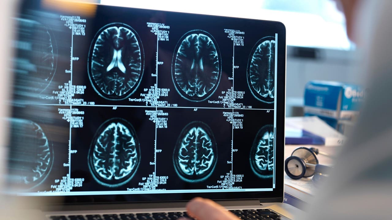
At Liv Hospital, we use MRI technology to see the brain’s details. This helps us find and treat many brain problems. MRI is safe and doesn’t hurt, using a big magnet, radio waves, and a computer to show us what’s inside without X-rays.
We’re all about being precise and caring for our patients. MRI lets us see how the brain is doing and find injuries. It’s a key tool for us to help you understand your brain health and recovery.

MRI technology has changed brain imaging a lot. It gives unparalleled visualization of soft tissues. This is something other imaging methods can’t do. We’ll look at how MRI works and how it makes detailed brain images.
MRI uses magnetic resonance to create images. It uses a strong magnetic field and radio waves. These align hydrogen nuclei in the body and then disturb them to produce signals. These signals are used to make detailed images.
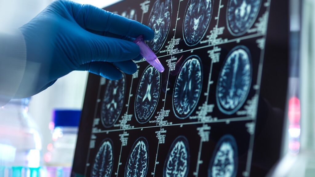
MRI technology captures signals from the brain’s tissues. These signals are then processed to make high-resolution images. These images show subtle details about brain structures and any abnormalities.
There are many MRI sequences for brain scans. T1-weighted, T2-weighted, and FLAIR sequences are some examples. Each sequence shows different information about brain tissues and possible problems. By choosing the right sequence, doctors can understand brain health better.
Knowing these basics is key to understanding MRI’s role in detecting brain damage and assessing brain activity. We’ll dive into this more in the next sections.
Understanding MRI’s role in diagnosing brain damage is key. MRI is a top tool for checking the brain. It shows detailed images of brain structures.
Standard MRI scans can spot many brain damage types. This includes bleeding, swelling, tissue loss, and trauma effects. Sensitivity and Specificity in Brain Injury Detection
MRI is very good at finding brain injuries. It can spot small changes in the brain. This is key for early diagnosis and better treatment.
MRI beats other imaging methods like CT scans in many ways. It shows soft tissue details without harmful radiation. This makes it safer for more scans.
While CT scans are fast and used in emergencies, MRI gives clearer images. For example, MRI can tell if a stroke is ischemic or hemorrhagic. This helps doctors choose the right treatment.
In summary, MRI is a top tool for finding brain damage. It’s very sensitive and specific. Its detailed images are essential for brain health checks and care.
MRI has been a big step forward in finding brain damage. It shows detailed images that help spot different kinds of brain damage. This includes damage to the frontal lobe. Doctors use these images to better diagnose and treat patients.
MRI is great at finding bleeding and hemorrhages in the brain. It shows where, how big, and how bad the bleeding is. Knowing this helps doctors decide the best treatment. For example, MRI for brain damage is key in emergencies.
Brain swelling and edema are serious issues that can come from injury or illness. MRI can see how much swelling there is and how it affects the brain. This info is vital for taking care of patients.
MRI can also check for tissue loss and atrophy, signs of long-term brain damage. By looking at how much tissue is lost, doctors can understand the damage’s extent. This is very important when looking at frontal lobe damage MRI results.
In summary, MRI is a key tool for seeing brain damage. It gives detailed views of brain injuries and conditions. This helps doctors diagnose and plan treatments better.
Advanced MRI techniques are key to spotting brain damage from trauma. Traumatic brain injuries (TBI) are tricky to diagnose. We use different MRI methods to see how bad the injury is and help decide treatment.
Diffusion Tensor Imaging (DTI) is a top-notch MRI method. It shows how well white matter tracts in the brain are doing. It’s great for finding axonal injuries, which are common in TBI. DTI tracks water molecule movement in nerve fibers to spot damage.
Susceptibility-Weighted Imaging (SWI) is another advanced MRI tool. It’s super good at finding tiny blood spots in the brain. SWI is key for spotting small signs of TBI that regular MRI can’t see.
For concussion patients, we use special MRI tests. These tests mix DTI, SWI, and functional MRI (fMRI). They help us see both the structure and function of the brain after injury.
| MRI Technique | Application in TBI | Key Benefits |
|---|---|---|
| Diffusion Tensor Imaging (DTI) | Assesses axonal injury | Provides detailed information on white matter tract integrity |
| Susceptibility-Weighted Imaging (SWI) | Detects microhemorrhages | Highly sensitive to small blood products |
| Functional MRI (fMRI) | Evaluates brain activity | Helps assess functional changes post-injury |
Using these advanced MRI methods helps us better diagnose and treat TBI. This leads to better care for our patients.
MRI is key in finding stroke and brain damage from blood vessels. It shows us the brain’s health in detail. This helps us make quick and right treatment choices.
MRI tells us if a stroke is new or old. Knowing this helps doctors choose the best treatment. New strokes look different on MRI than old ones.
MRI helps us figure out if a stroke is caused by a blockage or bleeding. Blockage strokes show up on special MRI scans. Bleeding strokes are seen because of blood on other scans.
Abnormal blood vessels, like aneurysms, have unique signs on MRI. These signs help us spot and treat these problems. This lowers the chance of future strokes.
Thanks to MRI, we understand strokes and brain damage better. An MRI of the brain gives us vital info. This info helps us treat patients better.
fMRI shows us how the brain works by looking at blood flow changes. It’s a non-invasive way to see which brain parts are active during different tasks. This helps us understand brain function better.
fMRI tracks brain activity by watching blood flow. Active brain areas need more oxygen, so blood flow increases. This change is what fMRI detects to show brain activity. It uses BOLD imaging, which spots the difference in oxygen-rich and oxygen-poor blood.
fMRI is key in studying the brain. It helps us see how brain areas work together for tasks like memory and language. It’s also used before surgery to avoid important brain spots. Plus, it helps us understand brain disorders by showing how they affect brain activity.
Understanding fMRI data is important. It shows how the brain works normally and how it changes in disease. For example, it can show how the brain adapts after a stroke.
| Condition | fMRI Findings | Clinical Implication |
|---|---|---|
| Stroke | Altered brain activity patterns, compensatory activation | Understanding recovery mechanisms |
| Schizophrenia | Abnormal connectivity between brain regions | Insights into disease pathology |
| Alzheimer’s Disease | Reduced activity in memory-related brain areas | Early diagnosis and monitoring |
As we learn more about fMRI, we understand the brain better. This knowledge helps us find new ways to diagnose and treat diseases.
MRI is key in studying neurodegenerative diseases. It gives us detailed views of brain issues. We use MRI to spot and track diseases like Alzheimer’s, multiple sclerosis, and Parkinson’s.
MRI finds signs of Alzheimer’s, like shrinking hippocampal areas and thinning cortices. These signs show how the disease is growing. Advanced MRI techniques let us see these small changes, helping doctors care for patients.
MRI is vital for finding lesions in the brain and spinal cord of those with multiple sclerosis. It tracks how the disease is doing and if treatments work. We watch how lesion numbers change over time. High-resolution MRI images help spot small lesions early, leading to better care.
MRI helps in Parkinson’s and other movement disorders by looking at structural changes. It’s not the main tool for diagnosing Parkinson’s but helps rule out other causes. We also use functional MRI to study brain activity linked to these disorders.
Using these advanced MRI methods, we learn more about neurodegenerative diseases. This knowledge improves patient care and treatment results.
Using MRI to check for frontal lobe damage needs special methods. These methods give us detailed views of the injury. We use advanced MRI techniques to look at the frontal lobe. This area is key for thinking, moving, and making decisions.
To spot frontal lobe damage, we use certain MRI sequences. High-resolution T1-weighted images show the body’s structure. FLAIR sequences help find lesions.
| MRI Sequence | Application |
|---|---|
| T1-weighted | Anatomical detail |
| FLAIR | Lesion detection |
| Diffusion Tensor Imaging (DTI) | White matter tract assessment |
Frontal lobe injuries can come from head trauma or blood vessel problems. MRI spots injuries like contusions, lacerations, and axonal injuries.
By looking at MRI results, we can link frontal lobe damage to changes in thinking and behavior. This link is key for making rehab plans.
Knowing how frontal lobe damage affects people helps doctors give better care. This leads to better results for patients.
It’s important to know what MRI can’t do when it comes to brain damage. MRI is a great tool for finding problems, but it’s not perfect. There are things that can stop it from showing brain damage.
When you get an MRI after an injury, how soon you get it matters. Sometimes, damage won’t show up right away. You might need another scan later to see how bad it is.
Some brain damage is too small for MRI to see. This is because MRI can only show so much detail. It might miss tiny or subtle injuries.
There are technical issues that can mess with MRI results. Things like movement or metal implants can cause problems. Also, some people can’t have an MRI because of certain implants or conditions.
Key limitations of MRI include:
Knowing these limits helps doctors understand MRI results better. This is key for creating good treatment plans for brain damage patients.
MRI is key in finding and checking brain damage and activity. It has grown a lot, helping us see brain damage, find head injuries, and spot blood vessel problems. MRI’s detailed brain pictures are vital for diagnosing and treating brain diseases.
Can MRI find brain damage? Absolutely yes. It shows how bad the injury is, helping doctors decide on treatments and how to help patients recover. Also, fMRI tracks brain activity by watching blood flow changes. This helps us see which parts of the brain are affected by disease or injury.
MRI technology will keep getting better, giving us even clearer views of brain damage and activity. New research and MRI methods will make it even better at finding problems. This means doctors can give patients better care, leading to better health outcomes.
Yes, MRI can show brain damage. It can see bleeding, swelling, and tissue loss. This helps doctors diagnose neurological conditions.
Yes, MRI can detect brain activity. It uses changes in blood flow to see how the brain works. This gives insights into brain function.
An MRI of the brain shows detailed images. It helps doctors diagnose conditions like brain damage, stroke, and neurodegenerative diseases.
Yes, MRI can show frontal lobe damage. It uses special techniques to see injury patterns in the frontal lobe.
MRI uses advanced techniques to find traumatic brain injury. It looks for axonal injuries and microhemorrhages.
Yes, MRI can tell the difference between ischemic and hemorrhagic stroke. It shows the type of damage and vascular issues.
MRI has some limits. It depends on when the MRI is done after an injury. It can miss microscopic damage. Also, technical issues or health reasons might affect the quality or availability of the images.
Yes, MRI can show brain activity patterns in neurodegenerative diseases. It helps understand brain function and dysfunction.
Yes, MRI is useful for diagnosing and managing neurodegenerative diseases. It uses imaging biomarkers and can detect lesions.
MRI is better than CT scans for some brain damage. It’s more sensitive to soft tissue injuries. Plus, it gives detailed images without radiation.
Subscribe to our e-newsletter to stay informed about the latest innovations in the world of health and exclusive offers!
WhatsApp us