Last Updated on November 27, 2025 by Bilal Hasdemir
Nuclear cardiology is a key tool for diagnosing heart issues. It uses noninvasive nuclear cardiology techniques to check the heart’s health and spot heart diseases.
These methods help doctors see how well the heart pumps blood. They also show where a heart attack might have happened. Pharmacological stress tests use specific agents to simulate exercise-like stress on the heart. This way, doctors can find heart problems without surgery.
The nuclear cardiology techniques are great for people who can’t do regular exercise tests. These advanced tools help doctors create specific plans to treat heart disease.
Key Takeaways
- Nuclear cardiology techniques are noninvasive diagnostic tools.
- These techniques assess myocardial blood flow and heart function.
- Pharmacological stress tests are used for patients who cannot exercise.
- Nuclear cardiology helps diagnose coronary artery disease and other heart conditions.
- These diagnostic tools enable targeted treatment plans for heart disease management.
The Evolution and Importance of Nuclear Cardiology

Nuclear cardiology has changed how we care for the heart. It gives us key insights into heart diseases. This field is essential for diagnosing heart issues, understanding how severe they are, and predicting how patients will do.
Definition and Scope of Nuclear Cardiac Imaging
Nuclear cardiac imaging uses tiny amounts of radioactive tracers to see the heart and its blood flow. It helps us check how well the heart is working and if it’s damaged. This method is used to find coronary artery disease, see if treatments work, and track how the disease changes over time.
Historical Development and Current Applications
Nuclear cardiology started in the 1970s with the first use of myocardial perfusion imaging. Thanks to new technology and medicines, it has grown a lot. Now, it includes SPECT and PET scans. These are key for finding and managing heart diseases.
| Technique | Application | Advantages |
| SPECT | Myocardial Perfusion Imaging | Wide availability, cost-effective |
| PET | Cardiac Function and Viability | High sensitivity, accurate quantification |
| CCTA | Coronary Artery Imaging | Detailed anatomy, non-invasive |
Myocardial Perfusion Imaging: Cornerstone of Nuclear Cardiology
Myocardial perfusion imaging is a key tool in nuclear cardiology. It shows how blood flows through the heart. This method helps find coronary artery disease and check how the heart works under stress.
Single-Photon Emission Computed Tomography (SPECT) Principles
Single-Photon Emission Computed Tomography (SPECT) is a main tool in myocardial perfusion imaging. It uses a tiny amount of radioactive tracer. This tracer goes to the heart muscle based on blood flow.
SPECT imaging shows where the tracer is, giving a 3D view of the heart’s blood flow. It helps spot problems in blood flow.
| SPECT Characteristics | Description |
| Tracer Used | Technetium-99m Sestamibi or Thallium-201 |
| Imaging Technique | Gamma camera rotation around the patient |
| Information Provided | Three-dimensional image of heart perfusion |
Positron Emission Tomography (PET) in Cardiac Assessment
Positron Emission Tomography (PET) is also vital in myocardial perfusion imaging. It gives clear and detailed images. PET uses special tracers to check blood flow in the heart.
“PET myocardial perfusion imaging has emerged as a highly accurate method for diagnosing coronary artery disease, with superior image quality compared to SPECT.”
Journal of Nuclear Cardiology
PET is great because it can measure blood flow and is very accurate. It’s best for people with heart disease or who think they might have it.
The CCTA Test: Advanced Coronary Imaging Technology
The CCTA test is a big step forward in heart imaging. It lets doctors see the heart’s arteries without surgery. This helps find heart disease early and plan treatments.
What is Coronary Computed Tomography Angiography?
Coronary Computed Tomography Angiography (CCTA) uses CT scans to see the heart and its blood vessels. It’s a safe way to check for heart disease. CCTA gives clear pictures of the heart’s arteries, spotting blockages and plaque.
To do a CCTA test, a CT scanner takes pictures of the heart. A special dye makes the arteries stand out. The test is done in a hospital or imaging center and is usually easy for patients.
Technical Aspects and Image Acquisition in CCTA
CCTA uses top-notch CT scanners for fast data and smart image making. These scanners take detailed pictures of the heart’s arteries. This helps doctors see heart disease clearly.
| Technical Aspect | Description |
| CT Scanner Technology | Advanced scanners with high-speed data acquisition |
| Image Reconstruction | Sophisticated algorithms for image processing |
| Contrast Agent | Used to enhance visibility of coronary arteries |
The way images are taken in CCTA aims to get the best pictures while keeping radiation low. Thanks to these tech advances, CCTA is a trusted way to look at heart disease.
Nuclear Stress Testing: Protocols and Methodologies
Nuclear stress testing uses radiotracers and stress agents to check blood flow to the heart. It’s a non-invasive test that helps diagnose coronary artery disease and see how the heart works under stress.
The test’s protocols can change based on the patient’s health and the test’s goals. It usually involves either exercise stress or pharmacological stress to mimic heart stress.
Exercise Stress Testing Procedures
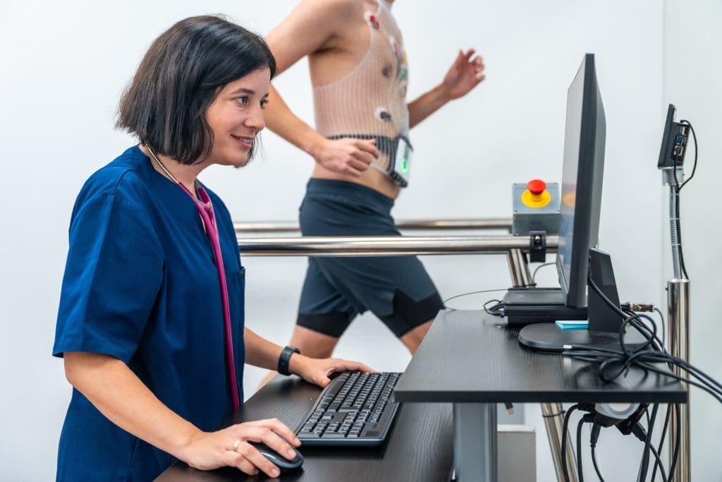
Exercise stress testing checks how the heart responds to physical stress. The patient walks on a treadmill or rides a bike while their heart rate and blood pressure are watched. The exercise gets harder to reach the target heart rate.
A radiotracer, like Technetium-99m Sestamibi (Cardiolite), is given at peak stress, and images are taken right after. This is done again at rest to compare stress and rest images for perfusion defects.
| Test Component | Description |
| Exercise Stress | Patient exercises on a treadmill or bike to achieve target heart rate |
| Radiotracer Injection | Technetium-99m Sestamibi is injected at peak stress |
| Imaging | Images are taken after stress and at rest to compare perfusion |
Cardiac Stress Test with Contrast: Benefits and Process
A cardiac stress test with contrast uses a contrast agent to better see the heart’s blood vessels. It gives detailed info on the heart’s structure and function, even under stress.
Using contrast in cardiac stress testing improves accuracy and helps see how severe coronary artery disease is. The process includes injecting the contrast agent during stress, then imaging with SPECT or PET.
“The use of contrast agents in cardiac stress testing has revolutionized the field of nuclear cardiology, enabling more precise diagnoses and tailored treatment plans.”
” Cardiologist
The images from the cardiac stress test with contrast are analyzed to spot any blood flow issues or perfusion defects. This helps guide further management and treatment plans.
Pharmacological Cardiac Stress Testing
Pharmacological cardiac stress testing is key when exercise is not possible. It helps check heart function and find coronary artery disease. It’s great for those who can’t do regular exercise tests because of health or mobility problems.
Lexiscan Nuclear Stress Test Procedure
The Lexiscan nuclear stress test uses regadenoson, a drug that acts like exercise on the heart. First, Lexiscan is given to the patient. Then, a radioactive tracer is injected. The heart’s reaction is captured by a gamma camera.
Key steps in the Lexiscan stress test procedure include:
- Preparation: Patients are told to avoid caffeine and certain meds before the test.
- Administration of Lexiscan: The drug is injected into the patient.
- Injection of radioactive tracer: A small amount of radioactive material is injected to see the heart’s blood flow.
- Imaging: A gamma camera takes pictures of the heart under stress and at rest.
Adenosine, Dobutamine, and Other Pharmacological Agents
Adenosine and dobutamine are also used in cardiac stress tests. Adenosine widens blood vessels, and dobutamine makes the heart work harder, like exercise. The right drug depends on the patient’s health and what the test aims to find.
Some key aspects of these agents include:
- Adenosine: It makes blood vessels wider, showing where blood flow is low.
- Dobutamine: It boosts heart rate and strength, good for those who can’t take adenosine or Lexiscan.
- Other agents: There are many other drugs used, each working in its own way.
Knowing about these drugs and their roles in heart tests is vital for good diagnosis and care.
Radiopharmaceuticals in Nuclear Cardiology
Radiopharmaceuticals are key in nuclear cardiology. They help diagnose coronary artery disease with advanced imaging. These substances go to the heart, showing how well it works.
Technetium-99m Sestamibi (Cardiolite) Applications
Technetium-99m sestamibi, known as Cardiolite, is a top choice in nuclear cardiology. It’s used for imaging the heart to spot coronary artery disease. It’s injected into the blood and goes to the heart muscle, showing where blood flow is low.
This agent offers clear images, is stable, and has low radiation. It’s great for checking if the heart muscle is working right and for finding heart disease in patients.
Thallium-201 and Newer Radioactive Tracers
Thallium-201 is used in nuclear cardiology for heart imaging. It’s been around for years to check heart muscle health. But, it has lower image quality and more radiation than technetium-99m sestamibi.
New tracers are being made to improve heart imaging and cut down radiation. Flurpiridaz is one example, promising better images and lower radiation. These new tracers aim to make nuclear cardiology safer and more accurate.
CCTA vs. Nuclear Stress Tests: Comparative Analysis
Two main tools are used to diagnose coronary artery disease: CCTA and nuclear stress tests. They help check the heart’s health but work in different ways. This affects how safe and accurate they are for patients.
Diagnostic Accuracy and Clinical Applications
CCTA shows detailed images of the heart’s arteries. It can spot blockages and plaque. Its accuracy is high, making it great for finding big problems in the arteries.
Nuclear stress tests, on the other hand, check how the heart works under stress. They use a radioactive tracer to see where blood flow is low.
Choosing between CCTA and nuclear stress tests depends on the patient’s situation. CCTA is good for those likely to have heart disease but can’t have invasive tests. Nuclear stress tests are better for checking how heart disease affects the heart’s function.
| Diagnostic Modality | Diagnostic Accuracy | Clinical Application | Radiation Exposure |
| CCTA | High for detecting stenosis | Anatomical assessment of coronary arteries | Moderate to High |
| Nuclear Stress Test | High for assessing ischemia | Functional assessment of coronary blood flow | Moderate |
Radiation Exposure and Safety Considerations
Both CCTA and nuclear stress tests use radiation, but doses are getting lower. CCTA usually has a higher dose than nuclear stress tests, but it can vary.
There are safety concerns with both tests. CCTA can harm kidneys in people with kidney problems and cause allergic reactions. Nuclear stress tests have risks from the radioactive tracer and stress agent.
Nuclear Stress Test vs. Angiogram: When to Use Each
It’s important to know the difference between nuclear stress tests and angiograms. This helps doctors choose the best test for patients with heart disease.
Nuclear stress tests and angiograms are used for different reasons. A nuclear stress test is not invasive and checks how the heart works under stress. It uses a radioactive tracer to see the heart muscle and blood flow. An angiogram, on the other hand, is invasive. It uses contrast material to see the inside of the heart’s arteries and find blockages.
Diagnostic Capabilities and Limitations
Nuclear stress tests can show if the heart is getting enough blood and if there’s damage. But they don’t show the heart’s arteries well. Angiograms, being invasive, can show the heart’s arteries in detail. They can find blockages. But, nuclear stress tests can’t find all heart problems, and angiograms have risks like bleeding.
- Nuclear stress tests are useful for:
- Checking blood flow to the heart
- Finding heart damage
- Being a safe first test
- Angiograms are useful for:
- Seeing the heart’s arteries clearly
- Finding blockages
- Helping with treatments like angioplasty
Invasive vs. Non-invasive Approaches to Coronary Assessment
Choosing between a nuclear stress test and an angiogram depends on the situation. Nuclear stress tests are often the first choice because they’re not invasive. Angiograms are used when there’s a strong chance of serious heart disease or when treatment is needed.
In summary, knowing what each test can and can’t do is key. Doctors use this knowledge to pick the right test for each patient. This way, they balance getting the needed information with keeping the patient safe.
CT Angiography: Advantages and Disadvantages
Coronary CT angiography (CCTA) is a cutting-edge imaging method. It gives detailed views of the coronary arteries, helping diagnose coronary artery disease. This non-invasive technique has changed how we diagnose heart issues, providing clear images of the heart’s blood vessels. It’s now a key tool for heart doctors.
Benefits of CT Angiography in Cardiac Diagnosis
One big advantage of CT angiography is its ability to show the coronary arteries clearly. It can spot blockages and other problems. This is great for people at risk of heart disease but without symptoms yet. The benefits of CCTA include being non-invasive, which lowers the risks of more invasive tests.
CT angiography also gives a full view of the heart’s structure. This helps doctors find issues early. A top cardiologist, says, “CCTA has changed how we diagnose and manage heart disease. It lets us see the heart’s blood vessels without surgery.”
Limitations and Possible Complications
Even with its benefits, CT angiography has limitations. One big concern is radiation exposure, but new tech has made doses much lower. Also, contrast dye can be a problem for some with kidney issues, so choosing the right patients is key.
There’s also a chance of getting false results, leading to more tests or false reassurance. So, it’s important to use CT angiography wisely and have experts interpret the results. Knowing both the advantages and disadvantages helps doctors decide when to use it for heart diagnosis.
Accuracy of Nuclear Cardiac Testing Methods
Nuclear heart stress tests are key in diagnosing heart issues. These tests help find coronary artery disease and how bad the blockages are.
Nuclear stress tests are very good at spotting coronary artery disease. How well they work depends on the test type, the patient’s health, and the doctor’s skill.
How Accurate is a Nuclear Heart Stress Test?
Nuclear heart stress tests are very accurate for finding coronary artery disease. They have a sensitivity of 80% to 90% and specificity of 70% to 90%. The test’s accuracy can change based on the patient’s age, sex, and health.
A study in the Journal of the American College of Cardiology showed the test’s sensitivity and specificity. It found the test was 87% sensitive and 75% specific in detecting coronary artery disease.
“Nuclear stress testing is a valuable tool for diagnosing coronary artery disease, with a high degree of accuracy and reliability.”
– Cardiologist
Does a Nuclear Stress Test Show Blocked Arteries?
A nuclear stress test can show if arteries are blocked, but it’s not a direct check. It looks at blood flow to the heart muscle under stress and at rest. If it finds a perfusion defect, it might mean there’s a blockage in a coronary artery.
The following table shows how well nuclear stress tests work in finding coronary artery disease:
| Test Type | Sensitivity | Specificity |
| Nuclear Stress Test | 80-90% | 70-90% |
| SPECT | 85-95% | 70-85% |
| PET | 90-95% | 85-95% |
In conclusion, nuclear cardiac testing methods, including nuclear stress tests, are very accurate. They help diagnose coronary artery disease and find out how severe the blockages are.
The SCOT-HEART Trial: Impact on Cardiac Imaging Practices
The SCOT-HEART trial marked a big change in how we diagnose coronary artery disease. It looked at how coronary computed tomography angiography (CCTA) helps find and treat this disease.
Study Design and Key Findings
The SCOT-HEART trial was a big study that compared two groups. One group got standard care, and the other got standard care plus CCTA. It included many patients with suspected heart disease.
It found that CCTA greatly improved diagnosis and changed treatment plans for patients.
Clinical Implications for CCTA Utilization
The trial’s results changed how we use cardiac imaging. More CCTA use means better diagnoses and more focused treatments. The table below shows how the trial’s findings changed patient care.
| Clinical Implication | Pre-CCTA | Post-CCTA |
| Diagnostic Accuracy | Limited by traditional methods | Improved with CCTA |
| Treatment Plans | Less targeted | More targeted and effective |
The SCOT-HEART trial has changed cardiac imaging forever. It shows how important CCTA is in today’s cardiology. Its results keep shaping clinical guidelines and practices.
Patient Experience During Nuclear Cardiac Procedures
Knowing what to expect during nuclear cardiac procedures can make a big difference. These tests, like nuclear stress tests, are key for checking heart health and spotting problems early.
Preparation and What to Expect During Testing
Getting ready is essential for a smooth nuclear cardiac procedure. Patients are often told to skip certain foods and meds beforehand. On test day, wear comfy clothes and be ready to stay calm on an exam table.
The test involves a tiny radioactive tracer in your blood. This helps the heart show up on images. You might do some exercise or take a special pill to stress your heart. A gamma camera captures pictures of your heart at work and at rest.
Managing Anxiety and Discomfort During Procedures
It’s vital to manage anxiety during these tests. Knowing what’s happening and why can help. Talking to your doctor or practicing relaxation can also be helpful.
Most people don’t feel much discomfort, but some might get anxious or feel trapped. Talking openly with the team can make you feel better.
Interpreting Nuclear Cardiology Results
It’s important for patients and doctors to understand nuclear cardiology test results. These tests show how well the heart works and if there’s coronary artery disease.
Understanding Perfusion Defects and Their Significance
Perfusion defects show where the heart’s blood flow is low. They can be reversible or fixed. Reversible ones mean the heart might get better with treatment. Fixed ones usually mean scar tissue from a heart attack.
- Reversible defects: Show ischemia, which might get better with treatment.
- Fixed defects: Usually mean scar tissue from a heart attack.
Abnormal Stress Test but Normal Catheterization: Explaining Discrepancies
Some patients have abnormal stress tests but normal catheterizations. This can happen for a few reasons. It might be due to microvascular disease or because each test works differently.
- Microvascular disease: Affects the heart’s small blood vessels.
- Diagnostic differences: Stress tests check heart function under stress. Catheterization looks at the coronary arteries directly.
Knowing about these differences helps doctors make better treatment plans.
Advanced and Hybrid Imaging Techniques
Nuclear cardiology is changing fast with new imaging methods. These advanced tools help doctors see the heart better. They give insights into how the heart works and its structure.
PET/CT and SPECT/CT Integration
PET/CT and SPECT/CT are changing nuclear cardiology. PET/CT shows how the heart uses energy and blood flow. SPECT/CT gives detailed views of blood flow and heart health. Together, they help doctors find and treat heart problems better.
Artificial Intelligence in Nuclear Cardiology
Artificial intelligence (AI) is becoming key in nuclear cardiology. It makes analyzing images faster and more accurate. AI finds patterns in data that humans might miss. This makes diagnosing heart issues quicker and more precise.
The future of nuclear cardiology is bright. It will keep improving with new imaging and AI. These advancements will help doctors diagnose and treat heart diseases better.
Conclusion: The Future of Nuclear Cardiology Techniques
Nuclear cardiology is growing, thanks to new imaging tech and techniques. These advancements help doctors diagnose better and care for patients more effectively. The future of nuclear cardiology looks bright, with ongoing research and development.
Coronary computed tomography angiography (CCTA) and other nuclear cardiology methods will shape clinical practice. Studies comparing CCTA with other tools, like nuclear stress tests, will guide doctors. This will help them choose the best ways to assess patients.
We can look forward to better diagnostic accuracy, less radiation, and more tailored treatments. Artificial intelligence and hybrid imaging will also boost nuclear cardiology. These advancements will help doctors provide better care for heart disease patients.
The future of nuclear cardiology is exciting, with new discoveries waiting. By keeping up with these advancements, doctors can offer better care for those with heart disease.
FAQ
What is a nuclear stress test, and how does it work?
A nuclear stress test checks how well the heart works when it’s stressed. This stress can come from exercise or medicine. A tiny bit of radioactive material is injected into the blood. This material builds up in the heart muscle, showing how well the heart is working.
What is the difference between a nuclear stress test and a CT angiogram?
A nuclear stress test looks at the heart’s function under stress. A CT angiogram, on the other hand, shows the coronary arteries and finds blockages. They are used for different reasons and in different situations.
How accurate is a nuclear heart stress test?
The accuracy of a nuclear heart stress test varies. It depends on the tracer used, the imaging quality, and the doctor’s skill. Generally, these tests are very good at finding heart disease.
Does a nuclear stress test show blocked arteries?
Yes, a nuclear stress test can show if arteries are blocked. It does this by looking at how well the heart gets blood. But it doesn’t show the blockages directly. It shows how the heart reacts to stress.
What is Lexiscan, and how is it used in nuclear stress testing?
Lexiscan, or regadenoson, is used for stress tests in people who can’t exercise. It makes the coronary arteries open up. This lets doctors see how well the heart is working.
What are the advantages and disadvantages of CT angiography?
CT angiography gives clear pictures of the coronary arteries. It’s great for finding blockages. But, it uses radiation and contrast agents. It also doesn’t show how well the heart is working.
What is the SCOT-HEART trial, and what were its key findings?
The SCOT-HEART trial compared CT angiography with usual care for heart disease. It found that CT angiography was better at finding problems. It also changed how doctors treated many patients.
How do I prepare for a nuclear cardiac procedure?
To prepare for a nuclear cardiac test, avoid certain medicines and don’t eat for a few hours. Remove any metal jewelry. Tell your doctor about any allergies or health issues.
What is the difference between SPECT and PET in nuclear cardiology?
SPECT and PET are both used in heart imaging. SPECT is more common for heart function tests. PET is better for detailed heart function and metabolism.
Can a nuclear stress test detect coronary artery disease?
Yes, a nuclear stress test can find coronary artery disease. It looks for areas where the heart doesn’t get enough blood. This can mean there’s a blockage.
What is the role of artificial intelligence in nuclear cardiology?
Artificial intelligence is becoming more important in heart imaging. It helps analyze images and predict outcomes. AI can make interpreting images faster and more accurate.


