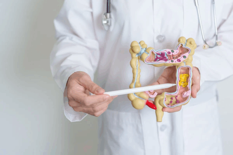
The proximal colon is a key part of the large intestine. It helps with digestion. It includes the cecum, ascending colon, and the start of the transverse colon. These parts are found on the right side of your abdomen.Explore the proximal colon — learn its anatomy, functions, and how it differs from other colon parts.
The large intestine is about 1.5 meters long. It’s wider and shorter than the small intestine. Knowing about the proximal colon helps doctors diagnose and treat problems.
Liv Hospital is a top place for care in the field of gastrointestinal health. They share important facts about the colon on the right side of the body.
Key Takeaways
- The proximal colon is a critical part of the large intestine.
- It includes the cecum, ascending colon, and proximal transverse colon.
- The proximal colon is located on the right side of the abdomen.
- Understanding its anatomy is vital for diagnosing related disorders.
- Liv Hospital provides expert insights into gastrointestinal health.
The Proximal Colon: Definition and Significance

The proximal colon is key for absorbing water and salts. It’s a vital part of the large intestine, essential for digestion.
What Constitutes the Proximal Colon
The proximal colon has three main parts: the cecum, the ascending colon, and the proximal transverse colon. Together, they help digest food.
- The cecum is the first part, getting contents from the ileum.
- The ascending colon absorbs more, moving contents up.
- The proximal transverse colon absorbs water and salts as contents move across the abdomen.
Role in the Digestive Process
The proximal colon is vital for absorbing water and salts. This solidifies stool and keeps electrolyte balance right.
The cecum gets contents from the ileum and continues absorption. The ileocecal valve makes sure contents move from the small intestine to the large one.
Key functions include:
- Absorption of water and electrolytes.
- Storage of digestive material.
- Processing of food residue.
Overview of the Right-Sided Colon
The proximal colon is on the right side of the abdomen, known as the right-sided colon. It includes the cecum, ascending colon, and part of the transverse colon.
This area is important for storing and processing food residue. Its right-sided position allows for specific relationships with other organs.
The right-sided colon is anatomically related to the right kidney and other surrounding structures.
Fact 1: The Proximal Colon Comprises Three Main Segments
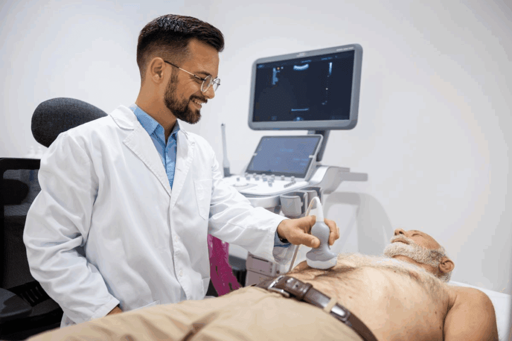
The proximal colon is split into three main parts. Each part has a key role in digestion. Together, they help move and process food.
The Cecum as the Initial Segment
The cecum is the first part of the proximal colon. It’s a sac-like structure that gets contents from the ileum. This is a key spot where the small and large intestines meet.
The cecum is found in the right lower part of the abdomen. It’s about 6-8 cm long. It holds digestive contents temporarily before they move on.
The Ascending Colon
The ascending colon is the second part. It goes up from the cecum along the right side of the abdomen. It ends at the hepatic flexure, where it turns into the transverse colon.
This part absorbs water and electrolytes from the digestive material. The ascending colon is behind the peritoneum, which lines the abdominal cavity.
The Proximal Transverse Colon
The proximal transverse colon is the third and last part. It spans the upper abdomen from the hepatic flexure to the splenic flexure. It’s the part of the transverse colon that’s closer to the start.
This segment is surrounded by the peritoneum and has a mesentery. This allows it to move more than the ascending colon. The proximal transverse colon also absorbs water and electrolytes.
Fact 2: Precise Location of the Proximal Colon in the Abdomen
Knowing where the proximal colon is is key for diagnosing and treating problems. It’s a big part of the large intestine. Its spot in the belly is important for how it works.
Right-Sided Positioning in the Abdominal Cavity
The proximal colon sits on the right side of the belly. It wraps around the small intestine on three sides. This right-sided positioning makes it different from other colon parts.
Its spot is not by chance. It’s placed just right for its job in digestion. Being on the right side lets it work with other important organs.
Anatomical Landmarks and Boundaries
The proximal colon has clear boundaries. It starts at the ileocecal valve and goes to the hepatic flexure.
- The cecum, the first part of the proximal colon, is in the right lower quadrant.
- The ascending colon goes up the right belly wall.
- The proximal transverse colon crosses from right to left, ending at the hepatic flexure.
Relationship to Surrounding Organs
The proximal colon is close to the liver, small intestine, and belly wall. Problems in the proximal colon can sometimes affect these nearby organs.
For example, the liver is near the hepatic flexure. Inflammation in the proximal colon can impact liver function. Knowing these connections is vital for good care.
Fact 3: The Cecum – Gateway to the Colon
The cecum is the first part of the colon. It connects to the ileum through a special valve. This is key for digestion, as it lets chyme move from the small intestine to the large one. The cecum is essential, starting the large intestine.
Structure and Dimensions of the Cecum
The cecum is about 6 cm (2.4 in) long. It gets the contents from the ileum. Its design includes the ileocecal valve, controlling the flow of digestive material. It’s a pouch-like part of the large intestine.
Connection to the Ileum via the Ileocecal Valve
The ileocecal valve is vital for controlling the flow from the ileum to the cecum. It stops backflow, making sure material moves right. This valve is a muscle, richly innervated and very specialized.
The Appendix and Its Attachment to the Cecum
The appendix is near the cecum’s junction with the ileum. Its exact role is debated, but it’s thought to aid in immunity. It’s a small, finger-like part attached to the cecum.
In summary, the cecum is a key structure, connecting the small and large intestines. Its unique shape and size help with digestion. Its connection to the appendix might suggest a role in immunity.
Fact 4: The Proximal Ascending Colon’s Unique Path
The proximal ascending colon is a key part of the large intestine. It runs vertically along the right side of the abdomen. This path is important for digestion and its connections with other parts of the body.
Vertical Trajectory Along the Right Abdominal Wall
The ascending colon starts at the cecum and goes up the right side of the abdomen. It doesn’t go straight but follows the shape of the abdominal wall. The part closest to the start is retroperitoneal, meaning it’s behind the lining of the abdomen.
As it goes up, the ascending colon is near the right kidney, liver, and muscles. Knowing this helps us understand how it works and where problems might happen.
Transition from Cecum to Hepatic Flexure
The ascending colon changes from the cecum to the hepatic flexure. The hepatic flexure is where the colon turns into the transverse colon. This change is important because it marks the end of the ascending colon.
“The hepatic flexure is an important anatomical landmark that can be a site of diverticulitis and tumors.”
Anatomical Relationships and Attachments
The proximal ascending colon is connected to the retroperitoneum by loose tissue. This lets it move a bit. It’s also tied to the fascia around the kidney and other nearby areas.
| Structure | Relationship to Proximal Ascending Colon |
| Right Kidney | Posterior and medial |
| Liver | Superior and anterior |
| Ileum | Connected via ileocecal valve to cecum |
Knowing these connections is key for surgeries and tests on the ascending colon.
Fact 5: The Proximal Transverse Colon’s Horizontal Course
The proximal transverse colon is a key part of the large intestine. It stretches from the hepatic flexure to the splenic flexure. This segment is vital for absorbing water and electrolytes.
Path from Hepatic to Splenic Flexure
The proximal transverse colon starts at the hepatic flexure. It then moves horizontally across the upper abdomen. It ends at the splenic flexure, turning into the descending colon.
Anatomical Support and Mobility
The proximal transverse colon is supported by the transverse mesocolon. This tissue connects the colon to the pancreas and other organs. It allows the colon to move freely while staying in place.
Distinguishing Features from Distal Transverse Colon
The proximal transverse colon is different from the distal part. Its location and blood supply are unique. The proximal part is more mobile and has a distinct blood supply.
| Characteristics | Proximal Transverse Colon | Distal Transverse Colon |
| Location | Extends from hepatic flexure to mid-transverse colon | Continues from mid-transverse colon to splenic flexure |
| Mobility | More mobile due to mesocolon attachment | Less mobile, transitioning towards descending colon |
| Blood Supply | Primarily supplied by middle colic artery | Receives supply from both middle and left colic arteries |
Knowing the unique features of the proximal transverse colon is key. It helps in diagnosing and treating colon-related issues. Its anatomy and function make it distinct from other parts of the GI tract.
Fact 6: Proximal vs. Distal Colon – Critical Differences
Knowing the differences between the proximal and distal colon is key for diagnosing and treating colon diseases. The large intestine is divided into four main parts: the cecum, colon, rectum, and anus. The proximal colon includes the cecum, ascending colon, and part of the transverse colon. The distal colon includes the descending and sigmoid colon.
Anatomical Distinctions Between Segments
The proximal and distal colon have unique features. The proximal colon is wider and has more sacculations than the distal colon. The cecum, the first part of the proximal colon, is a pouch-like structure. It receives contents from the ileum through the ileocecal valve.
- The ascending colon is behind the abdominal wall and goes up the right side.
- The proximal transverse colon is inside the abdomen and crosses from right to left. It is held up by the transverse mesocolon.
- The distal colon (descending and sigmoid colon) is narrower and more likely to have diverticular disease.
Functional and Physiological Differences
The proximal colon absorbs water, electrolytes, and some vitamins. It also ferments undigested carbs with the gut microbiota.
The distal colon stores and eliminates feces. The motility patterns are different. The proximal colon has more frequent, gentle contractions. The distal colon has stronger contractions to push feces towards the rectum.
Clinical Relevance of the Proximal-Distal Division
The difference between the proximal and distal colon is important for diagnosis and treatment. Diseases like colorectal cancer and diverticulitis affect these areas differently.
- Proximal colon cancers are often diagnosed later because of their location.
- Diverticulitis is more common in the distal colon, mainly in the sigmoid colon.
- Knowing these differences helps in targeted screening and treatment.
Fact 7: Blood Supply and Innervation Patterns
Understanding the blood supply and innervation of the proximal colon is key. It helps us see its role in digestion. The proximal colon, a big part of the large intestine, needs a strong blood supply and precise innervation to work well.
Arterial Supply from the Superior Mesenteric Artery
The proximal colon gets its blood mainly from the superior mesenteric artery (SMA). The SMA comes from the abdominal aorta. It supplies blood to the small intestine and a big part of the large intestine, including the proximal colon.
The SMA has branches that feed different parts of the proximal colon. These include:
- The ileocolic artery, which goes to the cecum and appendix.
- The right colic artery, which goes to the ascending colon.
- The middle colic artery, which goes to the transverse colon.
| Arterial Branch | Region Supplied |
| Ileocolic Artery | Cecum and Appendix |
| Right Colic Artery | Ascending Colon |
| Middle Colic Artery | Transverse Colon |
Venous Drainage Pathways
The venous drainage of the proximal colon follows a similar pattern to its arterial supply. The veins drain into the superior mesenteric vein (SMV). The SMV then joins the splenic vein to form the portal vein.
The SMV is key in the hepatic portal system. It carries nutrient-rich blood from the gut to the liver for processing.
Autonomic Nervous System Control
The proximal colon is controlled by the autonomic nervous system. This includes both sympathetic and parasympathetic parts. The parasympathetic part, mainly from the vagus nerve, helps with digestion.
The sympathetic part, from the sympathetic trunk, slows down digestion.
The balance between these two parts is important. It helps the proximal colon work right, controlling its movement, secretion, and blood flow.
Fact 8: Clinical Significance and Common Pathologies
The proximal colon is a key part of the large intestine. It faces many diseases and disorders. Its importance is shown by the common and serious problems it can have.
Proximal Colon-Specific Diseases
The proximal colon is at risk for several diseases, including:
- Diverticulosis: This is when small pouches form in the colon wall.
- Inflammatory Bowel Disease (IBD): Conditions like Crohn’s disease and ulcerative colitis can cause inflammation and damage in the proximal colon.
- Colorectal Cancer: The proximal colon is a common place for colorectal cancer to start, often from adenomatous polyps.
A study in a well-known medical journal said, “The proximal colon is a frequent site for colorectal cancer, highlighting the need for thorough screening and surveillance.”
“Colorectal cancer is one of the most common malignancies in the Western world, with a significant proportion arising in the proximal colon.”
Gastrointestinal Endoscopy, Volume 85, Issue 3, 2017
Diagnostic Approaches for Proximal Colon Issues
There are several ways to diagnose problems in the proximal colon:
| Diagnostic Method | Description | Advantages |
| Colonoscopy | Direct visualization of the colon using a flexible tube with a camera. | Allows for biopsy and removal of polyps. |
| CT Colonography | Radiological imaging of the colon using CT scans. | Less invasive than colonoscopy, can detect larger polyps and cancers. |
| Fecal Immunochemical Test (FIT) | A stool test that detects hidden blood. | Non-invasive, can be used for screening. |
Treatment Considerations and Challenges
Treatment for problems in the proximal colon varies based on the condition and its severity. For example, diverticulosis might be managed with diet changes and monitoring. On the other hand, colorectal cancer often needs surgery, possibly with chemotherapy or radiation therapy.
Dealing with diseases of the proximal colon is complex. It involves managing symptoms, treating the cause, and avoiding complications. As doctors face these challenges, early detection and tailored treatment plans are key.
Terminology Clarification: “Proximal” in Clinical Context
In medical talk, knowing what “proximal” means is key. It means something is closer to the start or a reference point. For the bowel, it means parts closer to the mouth.
Understanding “Proximal Bowel” Terminology
The term “proximal bowel” is important in medicine. It talks about the part of the intestine near the stomach. This includes the cecum, ascending colon, and part of the transverse colon. Knowing this helps doctors diagnose and treat problems in this area.
Distinguishing Proximal from Distal in Medical Communication
It’s important to know the difference between “proximal” and “distal”. “Proximal” means closer to the start, while “distal” means further away. For the colon, this helps doctors find where diseases or problems are. This guides their treatment choices.
Common Misconceptions About Proximal Colon Location
Many think the proximal colon is on the left side of the abdomen. But, it’s actually on the right side, including the cecum and ascending colon. Clearing up these mistakes is key for correct diagnosis and treatment.
In summary, understanding “proximal” in medical terms, like for the bowel and colon, is essential. It helps with clear communication, accurate diagnosis, and the right treatment plans.
Imaging and Assessment of the Proximal Colon
The proximal colon’s complex anatomy needs precise imaging for accurate diagnosis. It’s vital to see this area clearly to spot problems and plan treatment.
Colonoscopy Visualization and Challenges
Colonoscopy is key for seeing the proximal colon. It lets doctors directly look at the colon’s lining. This helps find polyps and other issues before they become serious.
“Colonoscopy remains the gold standard for detecting and removing precancerous polyps, significantly reducing the risk of colorectal cancer.”
Yet, colonoscopy faces challenges, mainly in the proximal colon. It’s hard to navigate and check every part well. Advancements in colonoscope technology have made it easier and clearer.
Radiological Imaging Techniques
Other imaging like CT colonography and barium enemas are also useful. They give detailed views of the colon. CT colonography is great for seeing the whole colon and finding other issues.
Choosing the right imaging depends on the patient, the suspected problem, and what’s available. Multidisciplinary collaboration between doctors is key to picking the best method.
Advanced Diagnostic Methods
New methods like capsule endoscopy and virtual colonoscopy are being looked into. They might offer new ways to see the proximal colon without being too invasive.
Artificial intelligence is also being used in imaging. AI can help spot problems and make diagnoses more accurate. As AI gets better, it will likely be more important for checking the proximal colon.
Conclusion: Maintaining Proximal Colon Health
Keeping the proximal colon healthy is key to feeling good. Eating foods high in fiber and avoiding processed foods helps a lot. Knowing how the proximal colon works helps us take care of it better.
Regular check-ups and managing health issues are important. They help avoid problems with the colon. By living healthily and knowing about the proximal colon, we can stay safe from colon issues.
Good health in the proximal colon means a healthy digestive system. It helps us absorb nutrients well and keeps diseases away. Taking care of the proximal colon is essential for a healthy body and mind.
FAQ
What is the proximal colon, and what are its main components?
The proximal colon is a key part of the large intestine. It includes the cecum, ascending colon, and the start of the transverse colon. It helps absorb water and salts during digestion.
Where is the proximal colon located in the abdomen?
You can find the proximal colon on the right side of your abdomen. It’s near the liver and small intestine, marked by the hepatic flexure.
What is the function of the cecum in the proximal colon?
The cecum is the first part of the colon. It connects to the ileum through the ileocecal valve. This valve controls the flow of food from the small intestine to the large intestine.
What is the difference between the proximal and distal colon?
The proximal colon includes the cecum, ascending colon, and the start of the transverse colon. The distal colon includes the descending and sigmoid colon. They have different roles and structures.
How is the proximal colon supplied with blood?
The proximal colon gets its blood from the superior mesenteric artery. This artery also supplies the small intestine.
What are the common pathologies associated with the proximal colon?
The proximal colon can get diseases like colon cancer, diverticulitis, and inflammatory bowel disease. Doctors use colonoscopy and imaging to diagnose these.
How is the proximal colon assessed and imaged?
Doctors use colonoscopy, radiology, and other methods to check the proximal colon. These help diagnose and treat problems.
What is the significance of maintaining proximal colon health?
Keeping the proximal colon healthy is important for your overall health. A good diet, regular check-ups, and managing diseases are key.
Is the cecum part of the colon?
Yes, the cecum is part of the colon. It’s the first pouch connected to the ileum, starting the proximal colon.
What is the role of the proximal transverse colon?
The proximal transverse colon is a horizontal part of the colon. It goes from the hepatic to splenic flexures. Its anatomy and function are unique compared to the distal transverse colon.
References
Andrews, S., et al. (2013). Gallstone size e related to the incidence of post-cholecystectomy retained bile duct stones. Surgery Journal, 5(3), 143-147. Retrieved from https://www.sciencedirect.com/science/article/pii/S1743919113000484


