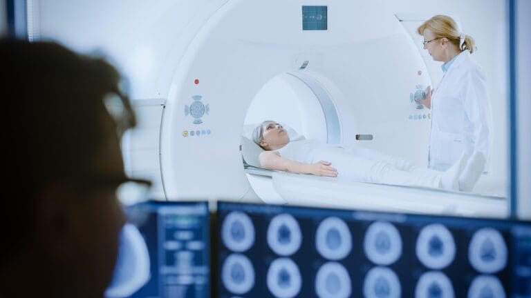
When brain MRI scans show abnormalities, patients want answers. At Liv Hospital, we offer a trusted, patient-focused way to understand these findings. We focus on T2 hyperintense foci, small spots that show up as abnormal areas.
T2 hyperintense lesions are common on MRI scans. Knowing what they mean is key for treatment. We explain how these spots show up on scans and what they might mean for your health. For more information, visit our partner site,Neurolaunch.

Key Takeaways
- T2 hyperintense foci are areas of increased signal intensity on T2-weighted MRI sequences.
- These lesions are often associated with aging, migraine, microvascular ischemic changes, or neurological conditions.
- The location of foci can hint at different possible causes or conditions.
- Age is a factor in T2 foci, with an increase often seen in older adults.
- FLAIR sequences are useful in detecting subtle changes in the brain.
The Basics of Brain Imaging and Abnormalities
Brain imaging is the cornerstone of diagnosing and managing neurological conditions. It allows specialists to visualize the brain’s structure and function, identifying abnormalities that may signal underlying disorders.

Common Brain Imaging Techniques
There are many brain imaging methods used in neurology. Each one gives us different info about the brain. Here are a few:
- Magnetic Resonance Imaging (MRI): MRI is a safe way to see the brain. It uses strong magnets and radio waves to show detailed images. It’s great for spotting soft tissue issues.
- Computed Tomography (CT) scans: CT scans use X-rays to make brain images. They’re fast and useful, often in urgent cases.
- Functional MRI (fMRI): fMRI tracks blood flow to show brain activity. It’s good for checking how the brain works.
These imaging methods are vital for finding and tracking brain problems. This includes those with T2 hyperintense lesions.
How Abnormalities Appear on Different Scans
Brain problems look different on different scans. It depends on the type of problem and the scan used. For example:
- T2-weighted MRI: T2 images spot water changes well. They’re great for finding swelling and lesions. T2 hyperintense lesions show up bright.
- FLAIR sequences: FLAIR sequences are good for finding brain lesions. They’re helpful for spotting problems near the brain’s center.
Knowing how scans show brain problems is key for correct diagnosis and treatment. By using many imaging types, we get a full picture of a patient’s health.
What Is Foci in the Brain? Definition and Characteristics
The term “foci” in neurology simply refers to specific, localized areas in the brain that appear distinct from the surrounding tissue on scans like an MRI. While the term can sound alarming, knowing the characteristics helps in accurate diagnosis.
Medical Definition of Brain Foci
Brain foci are distinct spots that can appear as hyperintense (bright) or hypointense (dark) areas depending on the MRI sequence. The texture, shape, and intensity of these spots provide critical diagnostic information.
We categorize brain foci based on their morphology and distribution. For instance, punctate foci are small, dot-like spots that can be scattered throughout the brain. Understanding the nature of these spots is the first step in evaluating their clinical impact.
How Foci Appear on Imaging Studies
On MRI scans, foci show up as areas that look different.Hyperintense T2 foci are bright, showing more water or other changes. Where these spots are found can help us understand what they might be.
We use different MRI sequences to learn more about foci. For example, FLAIR sequences help us tell apart lesions from other things like cerebrospinal fluid. By looking at how foci appear on different scans, we can start to figure out what they might mean.
It’s important to remember that foci don’t always mean a person has a disease. Sometimes, they show up in healthy people, like as they get older. But if foci are linked to symptoms or other scan findings, we need to look closer to understand their role.
T2 Hyperintense Foci Explained
T2 hyperintense foci are often seen on brain MRI scans. They are found using magnetic resonance imaging (MRI), mainly on T2-weighted sequences. Knowing what they mean is key for diagnosing and treating patients.
What Makes a Lesion “T2 Hyperintense”
A lesion is called “T2 hyperintense” if it looks brighter than the brain on T2-weighted MRI scans. This is because it has more water than normal brain tissue. T2-weighted images are great at showing tissue changes, helping spot many diseases.
These bright spots can be caused by inflammation, damage to the myelin sheath, lack of blood flow, or swelling. The exact reason can be guessed based on where the spot is, how big it is, and other details seen on the scan.
Common Locations of T2 Hyperintensities
T2 hyperintense spots can show up in different brain areas. Where they are can hint at what’s causing them. They often appear in the white matter, like near the ventricles or under the brain’s surface. These spots are more likely to be related to small blood vessel problems or damage to the myelin sheath.
| Location | Common Associations |
| Periventricular White Matter | Small vessel disease, demyelination |
| Subcortical White Matter | Small vessel disease, migraines |
| Basal Ganglia | Vascular disease, metabolic disorders |
To understand T2 hyperintense foci, we need to look at the whole picture. This includes the patient’s symptoms and sometimes more scans. These spots should always be seen in the context of the patient’s overall health.
Punctate T2 Hyperintensities: Small But Significant
Punctate T2 hyperintensities are small but important findings on MRI scans. They are tiny spots, just a few millimeters in size. These spots can show different neurological conditions.
Characteristics of Punctate Lesions
Punctate T2 hyperintensities are small and show up brightly on MRI scans. They are usually round or oval and found in the brain’s white matter. The significance of these lesions depends on their location, number, and the situation they are found in.
These spots are often found by accident during MRI scans for other reasons. But, they can sometimes be linked to certain neurological symptoms or conditions.
Differential Diagnosis for Punctate Findings
When punctate T2 hyperintensities are found, figuring out their cause is key. Possible reasons include:
- Small vessel disease
- Multiple sclerosis
- Migraine-related changes
- Age-related white matter changes
- Other demyelinating or inflammatory conditions
The patient’s age, medical history, and symptoms are important in figuring out the cause. More tests or follow-up scans might be needed to watch these spots and help decide how to manage them.
FLAIR Imaging and Hyperintense Foci Detection
FLAIR imaging has changed how we find hyperintense foci in the brain. We use FLAIR sequences to see lesions better, which is key for neurological conditions. This method is vital for spotting and tracking brain issues.

Advantages of FLAIR Sequences
FLAIR sequences bring many benefits for finding hyperintense foci.
Key benefits include:
- They make lesions stand out by suppressing cerebrospinal fluid (CSF) signal
- They help spot lesions near the ventricles or subarachnoid space
- They show white matter lesions more clearly
These perks make FLAIR sequences great for diagnosing conditions like multiple sclerosis and other demyelinating diseases.
Punctate FLAIR Hyperintense Foci Interpretation
Punctate FLAIR hyperintense foci are small, bright spots on FLAIR images. When we see these, we think about the patient’s situation and other imaging details. We look at several things, like:
- Where the lesions are and how they spread
- If there are other imaging signs
- The patient’s symptoms and medical history
Understanding these spots helps us figure out what’s going on in the brain. It helps us narrow down what might be wrong and how to treat it.
Scattered Foci of T2 Hyperintensity: Patterns and Distribution
Scattered T2 hyperintensities on MRI scans can show different health issues. It’s key to look at where these spots are. When we see scattered foci in brain scans, we need to know what it means for the patient.
What “Scattered” Pattern Indicates
A “scattered” pattern means these spots are spread out in the brain. This can happen in small vessel disease, demyelinating diseases, and normal aging. It shows the problem is not in one place but all over.
Scattered foci have a few main traits:
- Lesions are found in many parts of the brain.
- The size and shape of these spots vary.
- They can be in both white and gray matter, but white matter is more common.
Clinical Significance of Distribution Patterns
Scattered T2 hyperintensities are linked to many health issues and risks. For example:
- Small vessel disease: These spots might show long-term small vessel damage.
- Demyelinating diseases: Like multiple sclerosis, which often shows up as scattered spots in white matter.
- Normal aging: As we get older, some white matter changes are normal.
Knowing how and where these spots appear helps doctors make a diagnosis. They look at the patient’s history, symptoms, and other tests to understand what these spots mean.
Age Related Changes vs. Pathological Findings
It’s key to know the difference between age-related changes and actual diseases in the brain. When we look at brain scans, we must tell apart normal aging and possible diseases.
Normal Aging and White Matter Changes
As we age, our brains change, including white matter. White matter changes show up on MRI scans, like T2-weighted and FLAIR images. These changes appear as hyperintensities, which are brighter spots.
With age, we see more white matter hyperintensities, mainly near the ventricles and deep areas. These changes might come from:
- Less myelin
- Gliosis
- Small vessel disease
When to Be Concerned About T2 Hyperintensities
While some white matter changes are normal with age, others might signal a problem. We should worry about T2 hyperintensities if:
- They cover a lot of area and blend together
- Their size or number grows a lot over time
- They’re linked to other brain symptoms or signs
- They affect key brain areas for thinking or moving
It’s important to link T2 hyperintensities with the patient’s overall health. We should look at their age, medical history, and how they’re doing neurologically.
In summary, while age brings changes, we must carefully check T2 hyperintensities. This helps us tell normal aging from possible diseases. By doing this, we can give better diagnoses and care for our patients.
Microvascular Ischemic Changes and Small Foci
It’s important to understand microvascular ischemic changes to diagnose and manage small vessel disease. These changes are linked to small foci seen on brain scans. We’ll look at how small vessel disease shows up and discuss how to prevent it.
Small Vessel Disease Manifestations
Small vessel disease affects the brain’s small blood vessels, causing neurological symptoms. Microvascular ischemic changes are a key sign of this disease, showing up as small areas of ischemia or infarction. These can be spotted with MRI.
The symptoms of small vessel disease vary. Some people may experience memory loss, trouble walking, or bladder control issues. Others might not show symptoms but have changes seen on scans.
“The presence of microvascular ischemic changes on MRI is a significant predictor of stroke and dementia.” – Expert in Neurology
Risk Factors and Prevention
Several factors increase the risk of small vessel disease and microvascular ischemic changes. These include high blood pressure, diabetes, high cholesterol, and smoking. Managing these risk factors is key to stopping the disease from getting worse.
- Hypertension control through medication and lifestyle changes
- Diabetes management with diet, exercise, and medication
- Smoking cessation programs
- Lipid-lowering therapy
Prevention aims to lessen the impact of these risk factors. A healthy lifestyle and managing health conditions can lower the risk of small vessel disease and its related changes.
We suggest a detailed plan to manage risk factors. Regular check-ups and monitoring with healthcare providers are essential. This approach can help lessen the effects of small vessel disease and boost brain health.
Neurological Conditions Associated with Brain Foci
It’s important to understand how brain foci link to neurological conditions. This knowledge helps in making accurate diagnoses and planning treatments. Brain foci, seen as T2 hyperintensities on MRI scans, point to several neurological disorders.
Multiple Sclerosis and Demyelinating Diseases
Multiple sclerosis (MS) is a chronic disease where the body attacks the central nervous system. T2 hyperintense lesions in the brain’s white matter are a key sign of MS. These lesions are usually found near the ventricles and follow the veins.

Advanced imaging helps us spot and track these lesions. They are vital for diagnosing MS and checking its activity. The type and location of these lesions help tell MS apart from other demyelinating diseases.
Migraines and Their Relationship to Brain Foci
Migraines are a neurological disorder marked by severe headaches and other symptoms like nausea. Studies show people with migraines often have white matter lesions, seen as T2 hyperintensities on MRI.
These lesions might be linked to migraine’s causes, possibly showing vascular or inflammatory issues. But, the exact connection between migraines and brain foci is being studied further.
Other Neurological Conditions
Other conditions like small vessel disease, vasculitis, and infections can also show brain foci. These include various diseases that affect the central nervous system.
| Condition | Characteristics | Typical Locations |
| Multiple Sclerosis | Demyelinating lesions | Periventricular, juxtacortical |
| Migraines | White matter lesions | Subcortical, deep white matter |
| Small Vessel Disease | Lacunes, white matter hyperintensities | Deep gray matter, white matter |
Brain foci have big implications for patient care. Knowing the neurological conditions they are linked to is key to giving the best care.
Clinical Evaluation of Nonspecific Foci of T2 FLAIR
When nonspecific foci of T2 FLAIR are found, a detailed check is needed. These spots can be just random or linked to brain issues. So, a careful look is key.
Diagnostic Workup When Foci Are Discovered
The process to check nonspecific foci of T2 FLAIR is thorough. First, a full medical history is taken. This looks for things like high blood pressure, diabetes, or past migraines.
Then, a neurological exam is done. It checks for any specific brain problems.
Key parts of the check-up are:
- Looking at past scans to see if things have changed
- Doing blood tests to check for things like multiple sclerosis or inflammation
- Using extra scans, like special MRI images, to learn more about the spots
Follow-up Imaging Recommendations
How often to check nonspecific foci of T2 FLAIR depends on what they look like and the first check-up. For people who are fine and don’t have symptoms, a follow-up MRI might be suggested in 1-2 years. This is to see if anything has changed.
If the spots might be linked to a certain problem, like small blood vessel issues, the check-ups might be more often. For example, if it looks like a problem with the protective covering of nerve fibers, more tests might be needed more often.
The aim of these follow-ups is to watch how the spots change or get better. This helps us take the best care of patients with these spots, treating any real problems and reducing risks.
Management Approaches for Significant Brain Foci
Managing significant brain foci needs a detailed plan based on the cause. Finding these foci leads to a deep look to figure out why they are there.
First, we need to know what causes the brain foci. This means doing a full check-up. This includes looking at the patient’s history, doing a physical exam, and using imaging tests.
Treatment Options Based on Underlying Causes
The treatment for brain foci depends on why they are there. For example:
- Multiple Sclerosis: Doctors use special medicines to help manage symptoms and slow the disease.
- Microvascular Ischemic Changes: The goal is to control risk factors like high blood pressure, diabetes, and high cholesterol.
- Migraines: To lessen migraine attacks, doctors suggest treatments and changes in lifestyle.
It’s important to make a treatment plan that fits the patient. This means considering their diagnosis, health, and what they prefer.
Lifestyle Modifications to Address Risk Factors
Medical treatments are not the only answer. Changing your lifestyle is also key. Here are some tips:
- Dietary Changes: Eat more fruits, veggies, whole grains, and lean meats.
- Regular Exercise: Do some exercise to keep your heart healthy.
- Smoking Cessation: Quit smoking to lower your risk of heart problems.
- Stress Management: Try stress-reducing activities like meditation or yoga.
By making healthy lifestyle choices, you can help manage your brain foci. This can slow down the progress of related conditions.
We stress the need for a detailed and personal plan to manage brain foci. This plan should include the right medical care and lifestyle changes.
Conclusion: Understanding the Clinical Significance of Brain Foci
Finding foci in the brain or T2 hyperintense lesions on an MRI can be unsettling, but it is often a manageable part of one’s health journey. Whether these findings represent normal aging, mild microvascular changes, or a specific condition like MS, the key is accurate interpretation.
At Liv Hospital, our neurology and radiology teams work together to translate these complex images into clear, actionable care plans. By understanding the nature of these lesions, we can address root causes, manage risk factors, and ensure the best possible quality of life.
For more professional discussions, visit our Linkdin page.
FAQ
What are foci in the brain, and what do they signify?
Foci in the brain are small spots that show up on MRI scans. They can mean different things, like certain brain diseases. It depends on where they are and how they look.
What are T2 hyperintense lesions, and how do they appear on MRI scans?
T2 hyperintense lesions are bright spots on MRI scans. They often show inflammation or damage in the brain. MRI scans help us find and understand these spots.
What is the difference between punctate and scattered foci of T2 hyperintensity?
Punctate foci are tiny, single spots. Scattered foci are many spots spread out. Knowing the difference helps us figure out what’s going on in the brain.
What is the clinical significance of punctate FLAIR hyperintense foci?
Punctate FLAIR hyperintense foci are small, bright spots on FLAIR scans. They can mean different things, like small brain damage. We look at them along with the patient’s symptoms and other scans.
How do age-related changes affect the appearance of T2 hyperintensities on MRI scans?
As we get older, our brains can show T2 hyperintensities on scans. We try to tell if these are normal or not by looking at age and other health factors.
What is the relationship between microvascular ischemic changes and small foci?
Small foci on scans can be linked to tiny blood vessel problems. We look at risk factors like high blood pressure and diabetes. This helps us prevent future problems.
What neurological conditions are associated with brain foci?
Brain foci can be linked to conditions like multiple sclerosis and migraines. We talk about these connections and how to treat them.
How do we manage patients with nonspecific foci of T2 FLAIR?
For patients with unclear spots on scans, we do a full check-up. We also suggest follow-up scans. This helps us keep an eye on any changes.
What are the treatment options for significant brain foci?
Treatment for big brain spots depends on the cause. It might include medicine or lifestyle changes. We guide patients on managing their condition and reducing risks.
What is the significance of understanding T2 hyperintense lesions and brain foci?
Knowing about T2 hyperintense lesions and brain foci is key for treating patients. We focus on accurate scan interpretation and matching it with the patient’s health.
What does “punctate t2 hyperintense foci” mean?
Punctate T2 hyperintense foci are small, bright spots on MRI scans. They can point to issues like small blood vessel disease or brain damage.
Are scattered t2 hyperintensities a concern?
Scattered T2 hyperintensities might be a worry, depending on their location and the patient’s health. We look into their significance and suggest next steps.


