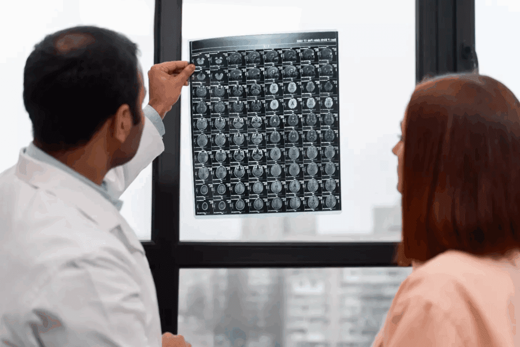
Seeing unusual dark spots on the brain in an MRI scan can worry patients and their families. At Liv Hospital, we know how critical accurate diagnosis and treatment are. Our team uses the latest medical knowledge to help with brain conditions.
Dark or black spots might show several health issues, like multiple sclerosis or small vessel disease. We aim to explain what these signs mean and how to manage them with new medical tools.
Key Takeaways
- Dark spots on the brain seen in MRI scans can indicate various neurological conditions.
- Accurate diagnosis is critical for effective treatment.
- Liv Hospital is committed to providing world-class healthcare with the latest evidence-based protocols.
- Our team of experts works closely with patients to understand their conditions and develop personalized treatment plans.
- Advanced MRI technology plays a vital role in diagnosing and managing neurological conditions.
Understanding Brain MRI Imaging

To understand brain MRI findings, we must first learn about MRI technology. Magnetic Resonance Imaging (MRI) is a non-invasive tool. It uses strong magnetic fields and radio waves to show detailed brain images.
How MRI Technology Works
MRI machines find signals from hydrogen atoms in the body. These signals come from the magnetic field and radio waves. They help create images of the brain’s structures.
The quality of MRI images depends on several things. These include the magnetic field strength, coil type, and sequence used. Knowing these factors helps us understand MRI scans better.
Different Types of Brain MRI Sequences
MRI sequences show different parts of the brain. There are several types:
- T1-weighted images, which show detailed anatomy
- T2-weighted images, which highlight changes in tissue water
- FLAIR (Fluid Attenuated Inversion Recovery) sequences, which hide fluid signals
- DWI (Diffusion-Weighted Imaging), which spots acute strokes
Each sequence gives unique insights into the brain. They help find black spots on the brain.
How Abnormalities Appear on MRI Scans
Abnormalities like dark spots show up differently on MRI scans. For example, inflammation or demyelination looks bright on T2-weighted images. Hemorrhages appear dark. Knowing how different problems look on MRI is key for correct diagnosis.
The look of black spots in brain MRI can mean many things. It could be something simple or serious. A radiologist’s accurate reading is vital to understand these findings.
Dark Spots on the Brain: Definition and Appearance

Dark spots on brain MRI scans can worry both patients and doctors. These spots, called hypointense lesions, show up as darker areas on MRI images.
What Are Hypointense Lesions
Hypointense lesions are darker than the brain tissue on MRI scans. They can mean tissue damage, disease, or other issues. The look of these spots changes with different MRI sequences.
MRI sequences like T1-weighted and T2-weighted images show different things about these lesions. For example, some spots might look dark on T1 images but bright on T2 images. This helps doctors understand what they are.
Common Terminology: Black Spots, Dark Spots, and Hypointensities
In medical reports, “black spots,” “dark spots,” and “hypointensities” mean the same thing. Knowing these terms helps doctors and patients understand MRI results.
The word “hypointense” means these spots show up as less intense on MRI scans. This term helps doctors describe these spots in a standard way.
General Significance of Finding Dark Spots
Dark spots on a brain MRI can mean serious health issues. They might be linked to multiple sclerosis, cerebrovascular disease, traumatic brain injuries, or neurodegenerative disorders.
Finding and understanding these spots is key for diagnosing and treating. More tests and doctor visits are often needed to figure out what they mean.
Multiple Sclerosis: A Primary Cause of Black Spots in Brain MRI
Multiple sclerosis (MS) is a major reason for black spots seen in brain MRI scans. It greatly affects patients’ lives. We will look into how MS leads to these black spots, known as lesions, and what they mean for the disease’s progress.
Characteristic MS Lesion Patterns
MS lesions in the brain show up as bright spots on T2-weighted MRI scans. They can appear dark on T1-weighted scans. These patterns help tell MS apart from other brain diseases. The lesions often appear in specific areas, which doctors check to diagnose MS.
A study on Medical News Today found that these lesions are a key sign of MS. Knowing about MS lesions is vital for correct diagnosis and treatment.
Chronic Active Lesions with Dark Rims
Chronic active MS lesions have a dark rim on MRI scans. These lesions show ongoing inflammation and damage. They are important because they show the disease is active, even if symptoms are not present. The presence of these lesions means the disease might be more aggressive, needing closer watch and possibly stronger treatment.
How MS Lesions Predict Disease Progression
The size, number, and activity of MS lesions on MRI scans can show how the disease will progress. A higher number of lesions often means a worse outcome. Chronic active lesions, as mentioned before, suggest a more severe disease. Watching these lesions helps doctors see if treatments are working and adjust them if needed.
MS is a leading cause of disability in young adults. Knowing about MS and its MRI signs is key to giving the best care. By studying black spots on brain MRI, we can manage MS better and improve patient results.
“The presence of chronic active lesions on MRI scans is a critical factor in determining the prognosis and treatment plan for MS patients.”
Cerebrovascular Causes of Dark Spots on Brain MRI
Dark spots on brain MRI scans often come from cerebrovascular diseases. These diseases harm the brain’s blood vessels, causing changes seen on MRI. We’ll look at small vessel disease, lacunar infarcts, and microbleeds as causes.
Small Vessel Disease and White Matter Changes
Small vessel disease harms the brain’s tiny blood vessels. It leads to white matter changes, which can show as dark spots on MRI. White matter changes are linked to aging, high blood pressure, and diabetes.
This disease can cause memory loss and raise stroke risk. It’s key to understand these changes to manage risk factors.
Lacunar Infarcts and Silent Strokes
Lacunar infarcts are small brain strokes caused by blocked arteries. They show up as dark spots on MRI, like on T1-weighted images. Silent strokes, including lacunar infarcts, might not show symptoms but can harm the brain over time.
Finding lacunar infarcts on MRI helps predict stroke risk and guide prevention.
Microbleeds and Hemosiderin Deposits
Microbleeds are tiny brain hemorrhages seen on MRI with susceptibility-weighted imaging (SWI). They look dark because of hemosiderin, a blood product. Microbleeds are linked to small vessel disease, high blood pressure, and amyloid angiopathy.
Microbleeds signal a higher risk of brain bleeding. They can also affect treatment choices for patients with atrial fibrillation.
Age-Related Black Spots on the Brain
Aging brings many changes to the brain, some of which show up as dark spots on MRI scans. These changes can be normal or signs of disease. It’s important to know the difference to make the right diagnosis and treatment.
Normal vs. Pathological Aging Changes
As we get older, our brains change in ways that can be seen on MRI scans. Some of these changes are just part of aging, while others might mean there’s a problem. For example, white matter hyperintensities are common in older adults and look like bright spots on MRI. But not all age-related changes are harmless; some can increase the risk of brain problems or diseases.
Research shows that some brain changes are more likely to be linked to serious aging issues. For instance, smoldering spots in the brain are linked to severe multiple sclerosis. This highlights the need to tell normal aging changes from those that are not.
White Matter Hyperintensities in Elderly Patients
White matter hyperintensities (WMH) are often seen in older adults on brain MRI scans. These lesions are thought to come from small vessel disease. They are linked to high blood pressure, diabetes, and atherosclerosis. WMH can be seen on T2-weighted and FLAIR MRI sequences, showing up as bright signals.
| Characteristics | Normal Aging | Pathological Aging |
| White Matter Hyperintensities | Mild, punctate lesions | Confluent, extensive lesions |
| Cognitive Impact | Minimal cognitive decline | Significant cognitive impairment |
| Associated Factors | Age, genetics | Hypertension, diabetes, atherosclerosis |
Cognitive Implications of Age-Related Spots
Age-related black spots on the brain, like white matter hyperintensities, are linked to cognitive decline in older adults. The impact on thinking varies with the severity and location of these spots. Studies show that those with more severe WMH are at higher risk of dementia and cognitive decline.
Understanding the effects of age-related brain changes on thinking is key to developing better care plans. By telling normal aging changes from those that are not, healthcare providers can offer more tailored care. This could help reduce the risk of cognitive decline in older adults.
Inflammatory and Infectious Origins of Brain Dark Spots
We look into how infections and inflammation can cause dark spots on brain MRI scans. These conditions can harm the brain, leading to lesions that show up as dark spots. Knowing about these issues is key for correct diagnosis and treatment.
Viral and Bacterial Encephalitis Patterns
Viral and bacterial encephalitis are serious infections that can inflame the brain. This can cause dark spots on MRI scans due to tissue damage and inflammation. Viral encephalitis, often from herpes simplex, can show up as necrotic lesions on T1-weighted images.
Bacterial encephalitis, from infections like tuberculosis, can also show dark spots. It may also cause abscesses.
The way these infections affect the brain can vary. For example, herpes simplex encephalitis usually hits the temporal lobes. Other viruses might target different areas. Spotting these patterns is key to figuring out the cause.
Autoimmune Encephalitis
Autoimmune encephalitis is when the immune system attacks the brain. Conditions like N-methyl-D-aspartate receptor (NMDAR) encephalitis can show up as dark spots on MRI. Diagnosing these conditions often involves clinical tests, lab work, and imaging.
The MRI signs of autoimmune encephalitis can vary. They might show up as widespread or focused changes. Spotting these changes is important for starting the right treatment.
Post-Infectious Demyelination Syndromes
Post-infectious demyelination syndromes, like acute disseminated encephalomyelitis (ADEM), happen after an infection. They cause the immune system to damage the myelin sheath around nerve fibers. ADEM can show up as multiple dark spots on MRI, mainly in the white matter.
It’s important to understand the clinical context and MRI findings for diagnosing these syndromes. The presence of multiple lesions and a recent infection are key clues.
Trauma-Related Black Spots in Brain MRI
MRI scans are key in spotting brain damage from injuries. They show dark spots that mean different kinds of damage. Knowing what these spots mean helps doctors treat patients better.
Acute vs. Chronic Traumatic Brain Injury Findings
Brain injuries can be either acute or chronic. Acute injuries show up as dark spots on MRI scans. These spots are due to swelling and bleeding.
Chronic injuries, on the other hand, might show up as smaller changes. These can include scarring or changes in brain tissue.
Diffuse Axonal Injury Characteristics
Diffuse axonal injury (DAI) is a common injury. It damages the brain’s axons. MRI scans show DAI as small dark spots, mainly in certain areas of the brain.
Evolution of Traumatic Lesions Over Time
Lesions from injuries change on MRI scans over time. At first, they might look dark because of bleeding. Later, they can look brighter as the body starts to heal.
In the end, these spots can leave behind signs that show up as dark spots on special MRI scans.
| Stage | MRI Sequence | Appearance |
| Acute | T2-weighted | Dark spots due to edema or hemorrhage |
| Subacute | T1-weighted | Hyperintense due to methemoglobin |
| Chronic | Gradient Echo | Dark spots due to hemosiderin deposits |
Migraine and Other Neurological Conditions Causing Dark Spots
Neurological disorders, like migraine, can show dark spots on MRI scans. This has led to more research into why this happens and what it means for patients. We will look into how these conditions affect brain scans and what they indicate.
White Matter Lesions Associated with Migraine
Migraine is linked to more white matter lesions on MRI scans. These lesions appear as bright spots on certain images and can be found in different brain areas. Research suggests these lesions might be connected to how often and severely migraines occur.
People with migraine, and those with aura, tend to have more of these lesions than others. It’s thought that the brain’s spreading depression during aura might cause these changes.
Neurodegenerative Disease Patterns
Neurodegenerative diseases can also show dark spots on MRI scans, with unique patterns. For example, multiple sclerosis shows lesions near the ventricles, while small vessel disease causes more widespread changes.
It’s important to understand these patterns for accurate diagnosis. Advanced MRI methods can give more details about these lesions.
Metabolic and Toxic Disorders
Metabolic and toxic disorders can also affect MRI scans. Vitamin B12 deficiency, for instance, can lead to white matter changes. Toxic exposures can cause different types of brain damage.
Doctors need to think about these causes when looking at MRI scans, mainly for patients with a history of metabolic or toxic issues. A detailed medical history and more tests can help figure out why dark spots appear.
Advanced Diagnostic Approaches for Black Spots on Brain MRI
Advanced diagnostic methods are key in understanding black spots on brain MRI. These techniques help us learn more about neurological conditions. They give us insights into the nature and importance of these spots.
Contrast-Enhanced MRI for Active Lesions
Contrast-enhanced MRI is great for finding active lesions. It uses a contrast agent to show inflammation or blood-brain barrier issues. This is very helpful in diagnosing conditions like multiple sclerosis.
“The use of contrast agents in MRI has changed how we find and track brain disease,” says a top neuroradiologist.
The contrast agent shows up in areas where the blood-brain barrier is broken. This makes active lesions stand out on MRI scans. It helps us diagnose and track treatment success over time.
Specialized Sequences: DWI, SWI, and FLAIR
Special MRI sequences like Diffusion-Weighted Imaging (DWI), Susceptibility-Weighted Imaging (SWI), and Fluid-Attenuated Inversion Recovery (FLAIR) give us more info on black spots.
- DWI shows how water molecules move and is good for finding acute strokes.
- SWI spots blood products and finds microbleeds or hemorrhages.
- FLAIR sequences help find brain lesions, even near CSF spaces.
Multimodal Imaging Approaches
Multimodal imaging combines different imaging methods for a fuller brain picture. It uses MRI with other scans like PET or CT. This helps us understand lesions better and their effect on brain function.
Multimodal imaging has many benefits. It improves diagnosis, disease understanding, and treatment planning.
“Multimodal imaging is the future of neurological diagnosis. By combining different imaging techniques, we can provide a more complete picture of the brain’s condition, leading to more effective treatment plans,” notes a specialist in neuroimaging.
Conclusion: What Patients Should Know About Brain MRI Findings
It’s important for patients to understand brain MRI findings. Dark spots on the brain can mean different things, like multiple sclerosis or age-related changes. Knowing this helps patients make better choices about their health.
Getting a correct diagnosis is vital. We’ve talked about the types of lesions and what they mean for treatment. For example, smouldering lesions in MS patients can lead to worse symptoms.
Learning about brain health is essential. Patients can make informed decisions by knowing about dark spots on MRI scans. This way, they can work better with their doctors to improve their health.
FAQ
What are dark spots on the brain seen in MRI scans?
Dark spots on the brain, seen in MRI scans, are called hypointense lesions. They look darker than the brain tissue around them. These spots can show different neurological conditions, like multiple sclerosis, small vessel disease, and brain injuries.
What is the significance of finding dark spots on the brain?
Finding dark spots on the brain means something different for everyone. It depends on the cause, where they are, and how many there are. We look at your medical history, do a clinical check-up, and use advanced imaging to figure out what they mean.
Can dark spots on the brain be a sign of multiple sclerosis?
Yes, dark spots can be a sign of multiple sclerosis (MS). MS lesions show up as dark spots on MRI scans, mainly in the brain’s white matter. We use special MRI sequences and clinical criteria to diagnose and track MS.
How do cerebrovascular causes lead to dark spots on brain MRI?
Cerebrovascular causes, like small vessel disease, can cause dark spots on MRI scans. These conditions damage the brain’s blood vessels, making changes visible on scans.
Are dark spots on the brain a normal part of aging?
Not all dark spots on the brain are just from aging. We check to see if they’re normal or a sign of a problem. This helps us tell the difference.
Can inflammatory and infectious conditions cause dark spots on the brain?
Yes, conditions like encephalitis and post-infectious demyelination syndromes can cause dark spots. We use advanced imaging and clinical checks to diagnose and treat these conditions.
How do traumatic brain injuries appear on MRI scans?
Traumatic brain injuries can show up as dark spots or other changes on MRI scans. The severity and type of injury determine what we see. MRI helps us understand the extent of the injury and track recovery.
Can migraine and other neurological conditions cause dark spots on the brain?
Yes, conditions like migraine can cause dark spots or white matter lesions on MRI scans. We look at these findings in the context of the patient’s overall health.
What advanced diagnostic approaches are used to evaluate black spots on brain MRI?
We use advanced methods like contrast-enhanced MRI and specialized sequences like DWI, SWI, and FLAIR. We also use multimodal imaging to understand black spots on brain MRI and find their cause.
How does Liv Hospital approach the diagnosis and treatment of conditions related to dark spots on the brain?
At Liv Hospital, we offer top-notch healthcare for conditions related to dark spots on the brain. Our team works together to make sure we diagnose and treat accurately and effectively.
References:
- MS brain lesions: causes, pictures, and MRI scans. Medical News Today. https://www.medicalnewstoday.com/articles/323976
- Differential diagnosis of tumor-like brain lesions. PMC. https://pmc.ncbi.nlm.nih.gov/articles/PMC10468256/
- Smoldering spots in the brain may signal severe MS. NIH News Releases. https://www.nih.gov/news-events/news-releases/smoldering-spots-brain-may-signal-severe-ms


