Last Updated on November 26, 2025 by Bilal Hasdemir

Discover why do I need an MRI scan for gallstones, what it shows, and how it compares to CT and X-ray. Your doctor might suggest an MRI scan for gallstones instead of CT or X-ray. At Liv Hospital, we use the latest imaging and care for the best results.
An MRI of the gallbladder uses strong magnetic fields and radio waves. It creates detailed images of the biliary system. This makes it great for spotting stones in the bile ducts that other tests might miss.
MRI scans show detailed images without using radiation. This makes them a key tool for finding and checking gallstones, even when other tests aren’t clear.

It’s important to know about gallstones to treat them well. They can cause many symptoms and problems. Gallstones are small, hard pieces that form in the gallbladder, a small organ under the liver.
Gallstones are made of cholesterol or bilirubin and can be different sizes. They usually happen when bile, made by the liver, gets too rich in cholesterol or bilirubin. This makes it harden into gallstones.
Types of Gallstones:
Gallstone disease can show itself in many ways, from mild to severe. Common symptoms include:
If gallstones are not treated, they can cause serious problems. These include inflammation of the gallbladder (cholecystitis), pancreatitis, or blockage of the bile ducts. MRI is key in finding these problems and helping with treatment.
| Complication | Description | Diagnostic Tool |
| Cholecystitis | Inflammation of the gallbladder, often caused by gallstones. | MRI, Ultrasound |
| Pancreatitis | Inflammation of the pancreas, potentially triggered by gallstones. | CT Scan, MRI |
| Bile Duct Obstruction | Blockage of the bile ducts, often by gallstones. | MRI, MRCP |
MRI is key in diagnosing gallstones. It gives us detailed images to understand the disease’s extent. We use MRI to check the gallbladder and bile ducts thoroughly. This is vital when first tests don’t show clear results or when we think there might be complications.
There are times when an MRI is needed to diagnose gallstones. This includes patients with suspected bile duct blockage, those with gallstone pancreatitis history, and those with unclear ultrasound results.
Specific scenarios where MRI is useful include:
Ultrasound is usually the first test for gallstones. But, sometimes it’s not clear or is affected by bowel gas or body shape. MRI is a good alternative in these cases.
MRI has better soft tissue contrast and can see bile ducts and surrounding areas better.
In complex cases, MRI has many advantages. It can spot bile duct stones, inflammation, and check for complications in surrounding tissues.
| Imaging Modality | Strengths | Limitations |
| Ultrasound | First-line, non-invasive, quick | Limited by bowel gas, operator-dependent |
| MRI | High soft tissue contrast, detailed bile duct visualization | Expensive, not as widely available |
| CT | Quick, widely available | Radiation exposure, less sensitive for non-calcified stones |
In conclusion, MRI is essential for diagnosing and managing gallstones, mainly in complex cases or when initial tests are unclear. It gives us detailed images of the gallbladder and bile ducts. This helps us make better decisions for patient care.
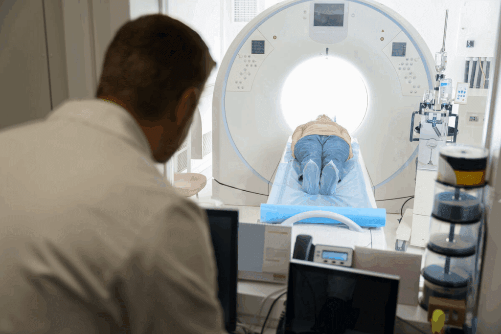
MRI technology has changed how we see the gallbladder and find gallstones. It uses strong magnetic fields and radio waves. This gives a clear view of the gallbladder and what’s around it.
MRI works on nuclear magnetic resonance. When a patient gets an MRI, they sit in a strong magnetic field. This field aligns hydrogen nuclei in their body.
Then, radio waves disturb these nuclei. This creates signals that make detailed images. MRI’s science is complex, using magnetic fields and radio waves for high-quality images.
MRI is great because it makes images without radiation. This is safer for patients who need many scans or are sensitive to radiation.
It uses special software to turn signals from the body’s hydrogen nuclei into detailed images. These images help doctors diagnose gallbladder problems.
| Imaging Modality | Radiation Use | Soft Tissue Detail |
| MRI | No | Excellent |
| CT Scan | Yes | Good |
| X-ray | Yes | Poor |
During a gallbladder MRI, patients lie in a big, cylindrical tube. It’s usually painless, but some might feel claustrophobia or discomfort from the machine’s noise.
To get better images, patients might get a contrast agent. This agent makes the gallbladder and its surroundings clearer.
The MRI is a key tool for seeing the gallbladder. It gives detailed images without radiation. This makes it a vital part of modern medical imaging.
For those with suspected gallstones or bile duct blockages, MRCP is often suggested. It’s a special MRI that shows the biliary and pancreatic ducts in detail. This helps in diagnosing and planning treatments for gallstone-related issues.
Magnetic Resonance Cholangiopancreatography, or MRCP, is a non-invasive test. It looks at the bile and pancreatic ducts. It uses a strong magnetic field and radio waves to show these areas clearly, helping spot blockages and stones.
MRCP is safe because it doesn’t need contrast agents injected into the ducts. This makes it more comfortable for patients.
Standard MRI scans show many internal organs. But MRCP focuses on the biliary and pancreatic ducts. It uses special sequences to highlight these ducts, making it easier to find problems.
MRCP stands out because it gives high-resolution images of the bile ducts without contrast agents. This makes it a key tool for diagnosing biliary system issues.
MRCP is better at showing the biliary system than other tests. This is key for spotting gallstones and bile duct blockages. It lets doctors see exactly where and what the problem is.
| Imaging Modality | Visualization of Biliary System | Use of Contrast Agents |
| MRCP | High-resolution images of bile ducts | No contrast agents required |
| Standard MRI | Comprehensive images of internal organs | May require contrast agents |
| CT Scan | Limited detail of bile ducts without contrast | Often requires contrast agents |
A study found MRCP to be very accurate for diagnosing bile duct issues. This shows its value in medical practice.
“MRCP has changed how we diagnose and manage biliary disorders. It’s a non-invasive and accurate way to see the biliary system.”
A leading gastroenterologist
MRI scans give a detailed look at gallstones and their complications. They often show things that other tests don’t. This advanced imaging is key for diagnosing complex gallstone cases.
MRI is great at finding stones in the bile ducts. These stones, called choledocholithiasis, can cause big problems if not treated right. MRI’s clear images help spot stones blocking bile flow.
MRI also spots inflammation and blockages from gallstones. It shows detailed images of the gallbladder and nearby tissues. This helps doctors diagnose conditions like cholecystitis and cholangitis.
MRI checks the tissues around gallstones too. It can find complications like abscesses or perforations that need quick action. MRI’s full view helps doctors make accurate diagnoses and plans.
MRI is very accurate in finding gallbladder stones. Studies show it’s as good as, or even better than, other tests. This accuracy is key for making the right treatment choices and improving patient outcomes.
In short, MRI is a big help in diagnosing gallstones and related issues. It can find bile duct stones, spot inflammation and blockages, and check surrounding tissues. This makes MRI a vital tool in managing gallstone disease.
CT scans have their own strengths and weaknesses when it comes to finding gallstones. They are not always the first choice but are important in some cases. This is true when looking for complications or when other tests can’t be used.
CT scans use X-rays to make detailed pictures of the body. This includes the gallbladder and bile ducts. The process involves moving an X-ray source and detectors around the patient. They capture many images to create a full view of the inside.
Key aspects of CT scan technology include:
Gallstones can be seen on CT scans, but it depends on their type. Calcified stones are easier to spot because they are denser. But, non-calcified or cholesterol stones are harder to find because they are less dense than the soft tissues around them.
“The sensitivity of CT scans for detecting gallstones varies, specially for non-calcified stones, highlighting the need for careful interpretation and possibly more imaging tests.”
— Expert in Radiology
Non-calcified gallstones are tricky for CT scans because they are similar in density to bile and soft tissues. Without enough contrast, these stones can be missed. This might lead to incorrect diagnoses or the need for more tests.
| Stone Type | Detectability on CT | Reason |
| Calcified | High | High density due to calcium |
| Non-Calcified | Low to Moderate | Similar density to surrounding tissues |
| Mixed | Moderate | Variable density |
Even though MRI is usually the top choice for gallbladder imaging, CT scans have their own benefits. They are better in emergency situations where quick action is needed. They are also good when MRI is not possible, like with certain metal implants.
Scenarios where CT might be preferred include:
X-rays are not the top choice for finding gallstones. They are used for many medical images, but they don’t work well for gallstones.
Most gallstones can’t be seen on an X-ray. This is because most stones are not made of calcium. Only a few stones are made of calcium, so they might show up on an X-ray.
Traditional X-rays don’t catch most gallstones. This is because most stones are made of cholesterol or other soft materials. So, doctors usually use other methods like ultrasound or MRI to find gallstones.
Even though X-rays have limits, there are other X-ray methods for looking at the bile ducts. ERCP uses an endoscope and X-rays to see inside. PTC injects a contrast agent into the bile ducts to show them on an X-ray.
Let’s look at how different imaging methods compare for finding gallstones:
| Imaging Technique | Effectiveness for Gallstones | Radiation Exposure |
| X-ray | Limited | Yes |
| Ultrasound | High | No |
| CT Scan | Moderate to High | Yes |
| MRI/MRCP | High | No |
In summary, X-rays are useful but not the best for finding gallstones. More advanced methods like ultrasound or MRI are better for spotting gallstones.
It’s important to know the strengths and weaknesses of MRI, CT, and X-ray for diagnosing gallstones. We look at their diagnostic value, including sensitivity, specificity, cost, and radiation exposure.
The ability of MRI, CT, and X-ray to detect gallstones varies.
The cost and how easy it is to get these tests can affect the choice of diagnostic tool.
| Imaging Modality | Cost | Accessibility |
| MRI | High | Moderate |
| CT | Moderate to High | High |
| X-ray | Low | High |
How much radiation each test uses is important, mainly for people who need more tests.
Choosing the right imaging test depends on the situation, the patient, and what’s suspected.
Healthcare providers can make better choices by considering these factors. This balances getting accurate diagnoses with keeping patients safe and accessible.
Advanced imaging is key in diagnosing and managing gallstones, even in tough cases. MRI is a top tool because it’s very sensitive and specific.
The role of advanced imaging in gallstone care is huge. MRI gives detailed pictures that help doctors make better treatment plans. This ensures patients get the best care possible.
When it comes to gallstones, picking the right imaging is vital. MRI is great because it shows the gallbladder and bile ducts without using harmful radiation. Using MRI helps us get better at diagnosing and treating gallstones.
As medical imaging gets better, MRI’s role in gallstone care will grow. We need to keep using advanced imaging to get the best results for patients with gallstones.
An MRI scan is recommended when other tests don’t give clear results. It helps us see the bile ducts and tissues around them. MRI is great because it shows detailed images without using harmful radiation.
Yes, CT scans can spot gallstones. But, they might miss stones that aren’t made of calcium. We often choose MRI for more detailed views, like when the situation is complex or we need to see the bile ducts.
Usually, X-rays can’t find most gallstones unless they’re made of calcium. We don’t usually use X-rays for gallstone diagnosis because they can’t show the gallbladder and bile ducts well.
MRI gives general pictures of the gallbladder and nearby areas. MRCP is a special MRI that focuses on the biliary system. It shows the bile ducts and gallbladder very clearly.
MRI uses a strong magnetic field and radio waves to make detailed pictures of the gallbladder and biliary system. It does this without using harmful radiation. This lets us look at the gallbladder and bile ducts from different angles.
MRI gives us important details about gallstones, bile ducts, and surrounding tissues in complex cases. This helps us make better treatment plans and manage any complications.
Yes, MRI is very good at finding gallstones, including those in the bile ducts. It can also spot problems like inflammation and blockages in the biliary system.
MRI is better than CT scans for finding non-calcified gallstones and checking the biliary system. But, CT scans might be chosen in emergency situations.
We usually suggest MRI for gallstone diagnosis because it’s very sensitive and doesn’t use radiation. But, the choice between MRI and CT scan depends on the situation and what the patient needs.
Yes, CT scans can show gallstones, but it depends on the stone’s type. Calcified stones are easier to see, but non-calcified ones might be missed.
X-rays can’t find most gallstones because they’re not made of calcium. We use more advanced tests like MRI or CT scans for accurate diagnosis.
MRCP, or Magnetic Resonance Cholangiopancreatography, is a special MRI that shows the biliary system in detail. We use it to check the bile ducts and gallbladder, mainly in complex cases or when we think there’s a blockage.
ShrEstha, G. (2023). Spigelian hernia: A rare case presentation and review of literature. Journal of Surgical Case Reports. Retrieved from https://www.sciencedirect.com/science/article/pii/S2210261223002079
Subscribe to our e-newsletter to stay informed about the latest innovations in the world of health and exclusive offers!